Figure 5. A novel in vitro assay to assess phagocytic activity in HESC-derived RPE cells. A: A schematic illustration of the novel in vitro retinal explant model system used to analyze phagocytosis. HESC-derived RPE
cells were cultured on a Matrigel™-coated filter and exposed to the photoreceptor side of human or pig retina placed on a
filter. The RPE-retina “sandwich” was cultured in HESC medium and held together by a plastic insert weighted down by the 6-well
plate lid. B: A Nomarski image of HESC-derived RPE exposed to a porcine retina explant for 48 h in vitro. The nuclei of cells are stained
with DAPI (blue). The outer nuclear layer (ONL), indicating the rod and cone nuclei, and inner nuclear layer (INL), specifying
the amacrine, bipolar, and horizontal cell layers are labeled. C-E: Immunohistochemical staining for rhodopsin in HESC-derived RPE cells exposed to porcine retina for 48 h in the novel retinal
explant system. C is a confocal projection and D-E are single confocal slices (<0.8 μm) merged with the Nomarski image. The rhodopsin staining (TRITC labeled) observed within
pigmented HESC-derived RPE cells is indicative of POS phagocytosis. The nucleus is stained with DAPI (blue). Scale bar in
B equals 50 µm; scale bar in C, D, and E equals 20 µm.
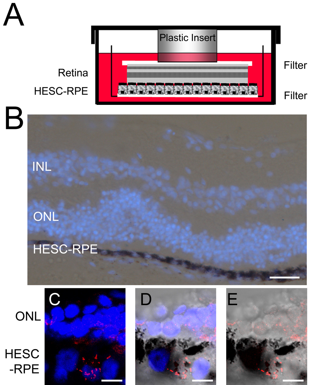
 Figure 5 of
Carr, Mol Vis 2009; 15:283-295.
Figure 5 of
Carr, Mol Vis 2009; 15:283-295.