Figure 3. HESC-RPE cells require MERTK for
the internalization of POS. HESC-RPE cells express the phagocytic
proteins (A) MERTK and (B) αVβ5 integrin. To determine
the importance of MERTK in the phagocytosis of POS, we treated HESC-RPE
cells with control IgG (C and F) or MERTK antibody (D
and G) for 1 h before exposure to Alexa Fluor® 488-labeled
porcine POS (C and D) or fluorescent polystyrene beads (F
and G) for 5 h. Cells were treated with trypan blue, washed,
and fixed. The number of internalized POS (E) and fluorescent
beads (H) was then quantified per field view (150 μm × 150 μm).
Data shown are mean±SEM (n=6). Pre-incubation with MERTK antibody had a
significant effect on the number of outer segments ingested by HESC-RPE
cells (p<0.01, Student t-test, n=6). There was no effect of
MERTK antibody on the ingestion of polystyrene beads by the cells
(p>0.05, Student t-test, n=6). Photomicrographs are single
confocal optical slices merged with Nomarski image (<0.8 μm). Scale
bars equal 50 µm. MERTK is labeled with Cy5 and counterstained with
DAPI, αVβ5 is labeled with FITC.
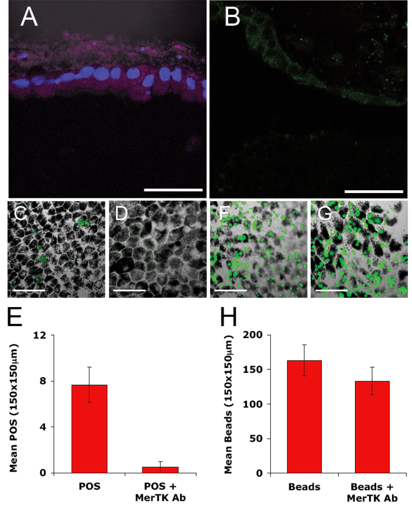
 Figure 3 of Carr, Mol Vis 2009; 15:283-295.