Figure 2. Increasing numbers of
fluorescently labeled porcine POS are phagocytosed by HESC-derived RPE
over time. A monolayer of HESC-derived RPE cells was exposed to Alexa
Fluor® 488-labeled porcine POS, treated with trypan blue to remove
fluorescent non-internalized POS, washed, fixed, and processed for
immunocytochemistry. Representative micrographs are shown from (A)
1 h and (B) 20 h together with respective Nomarski images (C
and D). Alexa Fluor® 488-labeled POS are in green. Scale bars
equal 50 μm. E Increased internalization of POS was observed
over time. Data shown are mean±SEM p<0.001, One-way ANOVA with
Bonferroni multicomparison test, (n=5); 0.5 h versus 4 h p<0.05, 0.5
h versus 20 h p<0.001, 1 h versus 20 h p<0.001, 4 h versus 20 h
p<0.05.
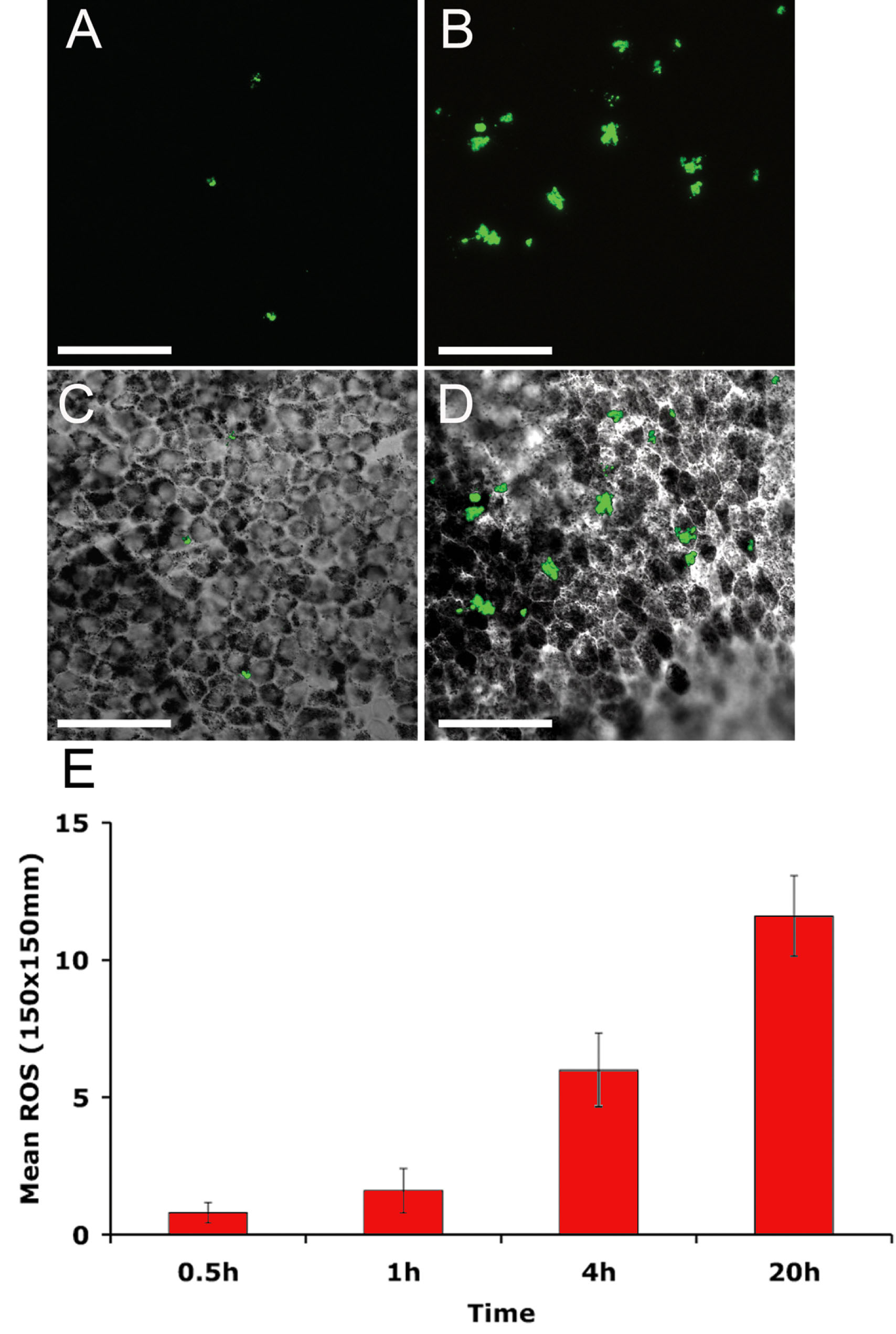
 Figure 2 of Carr, Mol Vis 2009; 15:283-295.