Figure 1. HESC-derived RPE cells
internalize fluorescently labeled POS isolated from the porcine eye. A:
The pigmented monolayer of HESC-derived RPE binds labeled POS (green). B:
A Nomarski image showing pigmented HESC-derived RPE overlaid with Alexa
Fluor® 488-labeled porcine POS. Evidence of internalization of outer
segments is clear by 4 h. C-D: Alexa Fluor® 488-labeled porcine
POS are associated with the apical surface of HESC-derived RPE
(delineated by TRITC-labeled Na+/K+ ATPase
immunostaining). D-E: POS are ingested by the cells and are
located close to the HESC-derived RPE nuclei (DAPI, stained blue).
Photomicrographs in C-E are single (y-axis) confocal optical
slices (<0.8 μm) from 3 separate HESC-RPE samples. In A-B,
scale bars equal 150 μm; in C-E, scale bars equal 10 μm.
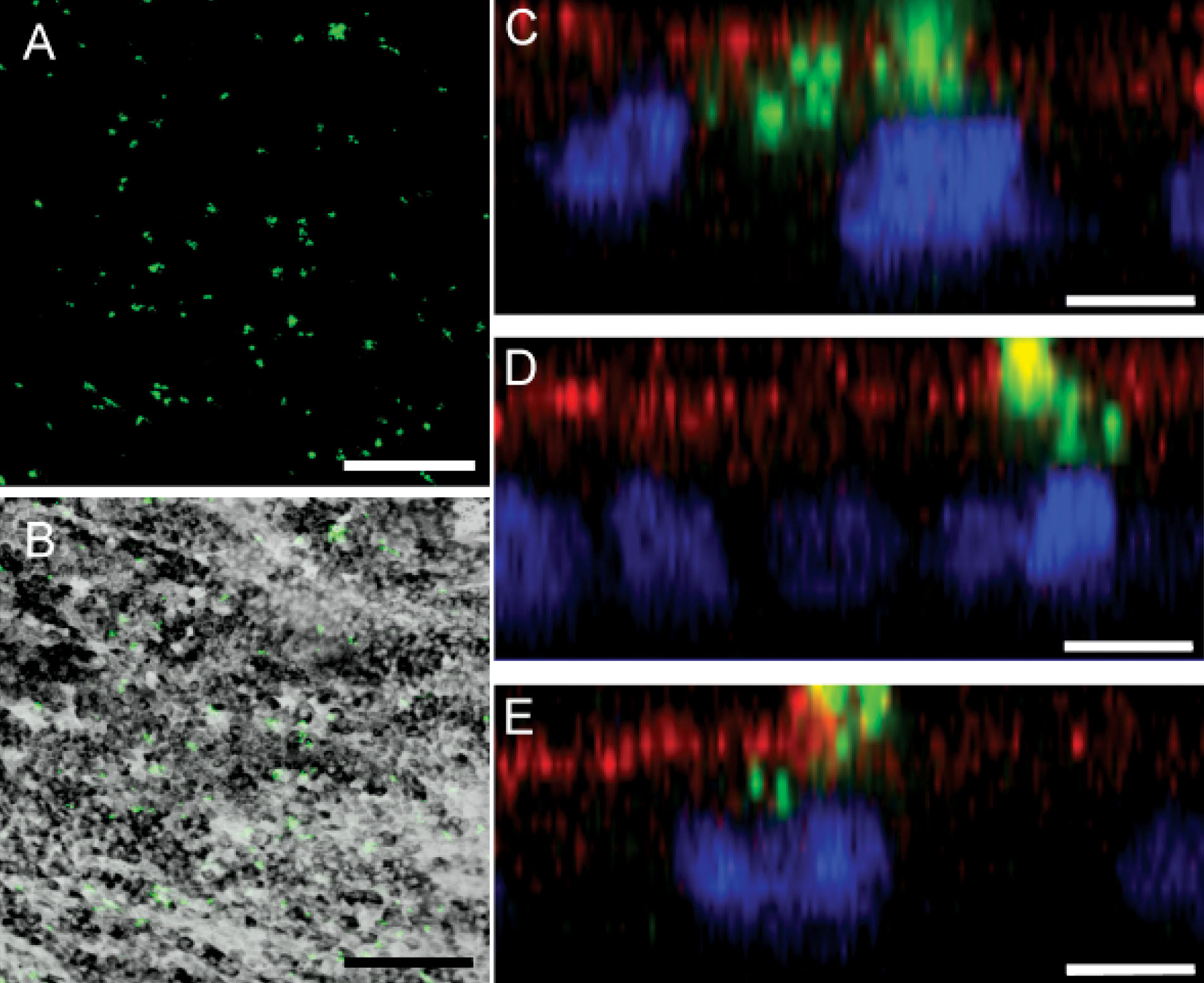
 Figure 1 of Carr, Mol Vis 2009; 15:283-295.