Figure 8. Fluorescent immunoreactivity of vimentin and GFAP in control and glaucomatous retinas. Panels A-D depict vimentin (red) antibody expression in control (A-B) and glaucomatous retinas after one week (C), and five weeks (D) of elevated IOP. Panels E-H indicate GFAP (green) antibody expression in astrocytes in control (E-F), one week (G), and five weeks of elevated IOP (H). Nuclei of the cells were marked with DAPI (blue). Abbreviations: ganglion cell layer (GCL), green fibrillary acidic protein
(GFAP), inner nuclear layer (INL), intraocular pressure (IOP), inner plexiform layer (IPL), outer nuclear layer (ONL), outer
plexiform layer (OPL). Scale bar represents 20 μm.
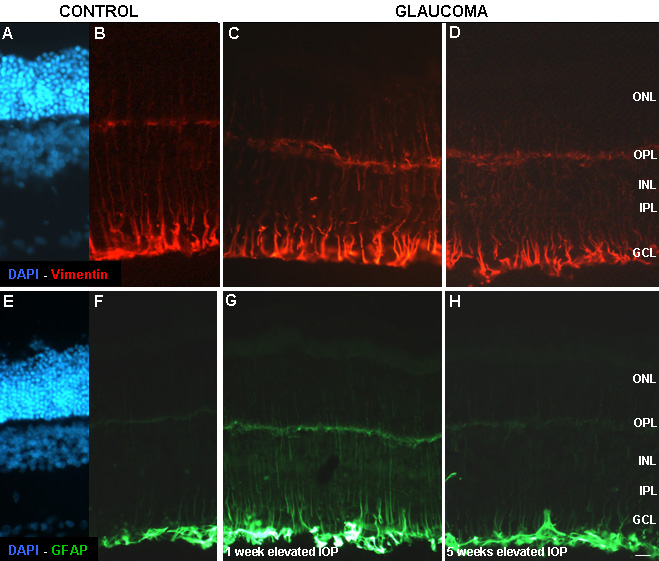
 Figure 8 of
Hernandez, Mol Vis 2009; 15:2696-2709.
Figure 8 of
Hernandez, Mol Vis 2009; 15:2696-2709.