Figure 2. M/L and S cone opsin immunohistochemical localization. Control (A, D, and E) and glaucomatous retinas (B, C, F, and G) were labeled with M/L and S cone opsin. Photographs B and F represent retinas after 1 week of elevated IOP and photographs C and G show retinas after 5 weeks of elevated IOP. Panel H represents the number of cone OS/mm in control and experimental groups as indicated (n=40 in all experimental groups). Abbreviations:
inner nuclear layer (INL), intraocular pressure (IOP), inner segment (IS), outer nuclear layer (ONL), outer plexiform layer
(OPL), outer segment (OS), Differential Interference Contrast (DIC), 4,6-diaminodiphenyl-2-phenylindole (DAPI). Scale bar
equals 20 μm.
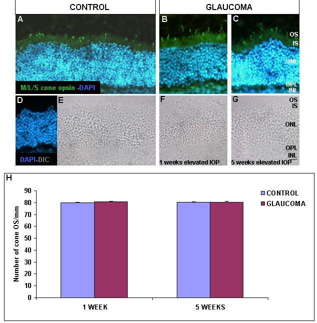
 Figure 2 of
Hernandez, Mol Vis 2009; 15:2696-2709.
Figure 2 of
Hernandez, Mol Vis 2009; 15:2696-2709.