Figure 6. Confocal images of p27 and PKCα
colocaliztion in RPE cells. PKCα and p27 have obvious
cytoplasmic localizations and slight nuclear localization, and mostly
colocalized in the cytoplasm of the cells stimulated with PMA,
thymeleatoxin, and siRNA- PKCα. PKCα (FITC, green
label) p27 (Cy3, red label), nuclei (Hotchest 33342, blue label), PKCα,
and
p27 colocalization (yellow label).
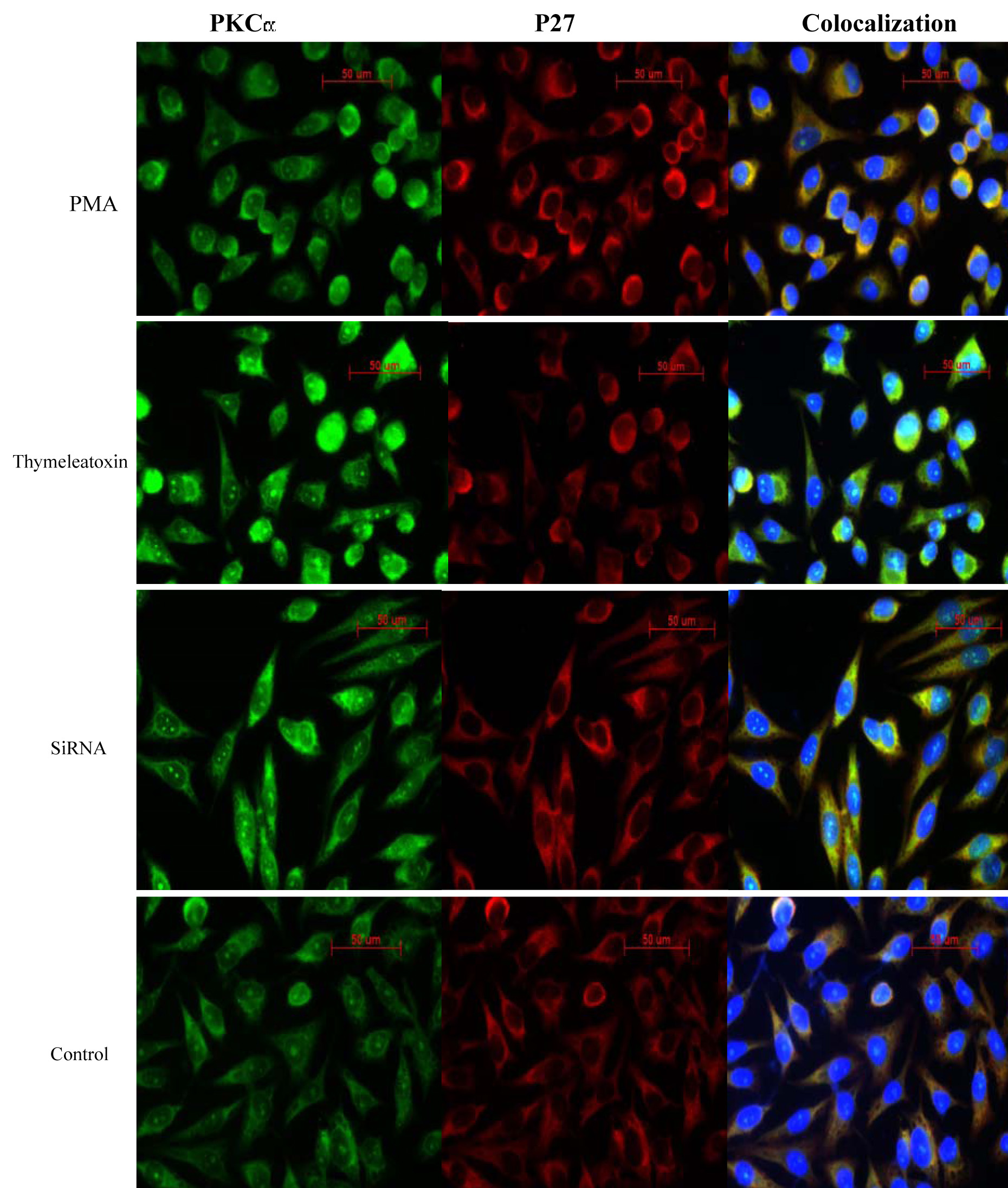
 Figure 6 of Gao, Mol Vis 2009; 15:2683-2695.