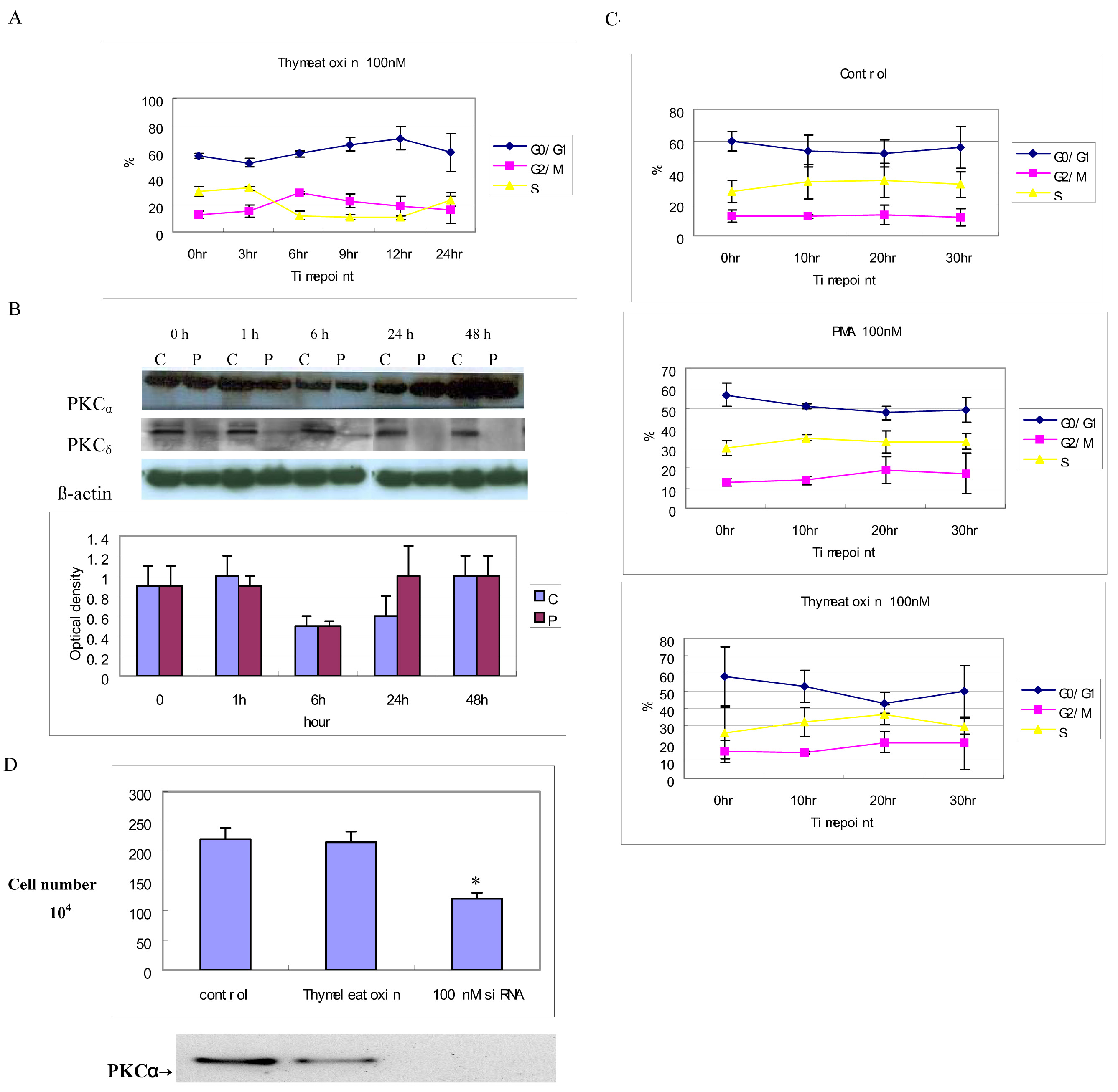
Figure 3. PKCα is necessary and
sufficient to affect cell cycle progression. A: Flow cytometry
analysis of RPE cells after 100 nM thymeleatoxin treatment shows a cell
cycle progression profile similar to that obtained with PMA in eight
experiments. B: western blot analysis shows that PKCα
was rapidly translocated to the membrane by thymeleatoxin and
downregulated within 24 h, the protein remained undetectable after 48 h
of treatment, however, PKCδ was not translocated and was not
downregulated at all time points. Eighty micrograms of protein was
loaded in each well. Optical density of PKCα determined by
densitometric imaging is shown (Mean±SD, n=3). The β-actin band with
42 kDa is used for quantitation. C: Flow cytometry
analysis of RPE cells shows that there was no significant change in the
cell cycle progression following PMA or thymeleatoxin restimulation
when compared with the control over the 30 h time course after 48 h of
PMA treatment. D: PKCα activity regulates the growth
rate of RPE cells. Approximately 110,000 RPE cells were seeded and then
incubated with thymeleatoxin or siRNA-PKCα for 24h. The
numbers of cells were counted using a Coulter Counter and displayed in
the top panel (* p<0.0001). Western blot using an anti-PKCα
antibody showed that the total PKCα level was dramatically
decreased in siRNA-PKCα treated cells; 40 µg of protein was
loaded in each well.

 Figure 3 of Gao, Mol Vis 2009; 15:2683-2695.
  Figure 3 of Gao, Mol Vis 2009; 15:2683-2695.