Figure 6. Localization of ephrin-A1 (A-D) and -A4 (E-H, K-M) in human and macaque retinas. A: Low magnification, dark field image of a section from a 13 WG human retina showing ephrin-A1 mRNA expression (white) in cells distributed throughout the optic nerve head and immediately adjacent retina. The yellow
box indicates the region shown in bright-field illumination (left inset). A triangular cluster of cells expressing ephrin-A1 mRNA in the outer neuroblastic layer of the retina, adjacent to the optic nerve is denoted by a heavy arrow. These cells
were also immunoreactive for Pax2 (see F and G). Ephrin-A1 expressing cells in the optic nerve head and adjacent to Bergmeister's papilla are indicated by the white box. The inset
to the right shows colocalization of ephrin-A1 (white) and Pax2 (green) in these cells. The far right inset shows cells in the same regions of the adjacent sense-treated section. B-D: High magnification image of the optic nerve in a Fd 55 macaque shows ephrin-A1 immunoreactivity (green) on the plasma membrane of cells (arrows) whose nuclei were Pax2 immunoreactive (red), indicating the cells are astrocytes. Inset in D shows a high magnification view of an astrocyte with punctate-like ephrin-A1 immunolabeling on its plasma membrane. E-G: Dark field scan of the optic nerve head showing ephrin-A4 mRNA expression (white) in Pax2-positive astrocytic cells (green) in the 13 WG human retina. H: The ephrin-A4 sense probe showed no specific labeling in a similar field in the optic nerve head. J: Goat IgG, control serum for ephrin-A1 and -A4 (Alexa 488), did not label astrocytes in Fd 55 tissue. K: At Fd 55, ephrin-A4 immunoreactivity (green) was detected in the optic nerve head (ONH) and adjacent retina. L-M: Double labeling with anti-Pax2 (red) shows ephrin-A4 immunoreactivity surrounding the Pax2 immunoreactive nuclei, indicating they are astrocytes (arrowheads). Astrocytes in the outer neuroblastic layer at the retina-optic
nerve junction are Pax2 immunoreactive (large arrow), and cells on the outer margin of this population are also ephrin-A4 immunoreactive. Scale bars in A represent 50 µm. Scale bars in panels B-M represent 25 µm. Abbreviations: antisense (AS); ganglion cell layer (GCL); immunoreactivity (IR); nerve fiber layer (NFL);
outer neuroblastic layer (ONB); optic nerve head (ONH).
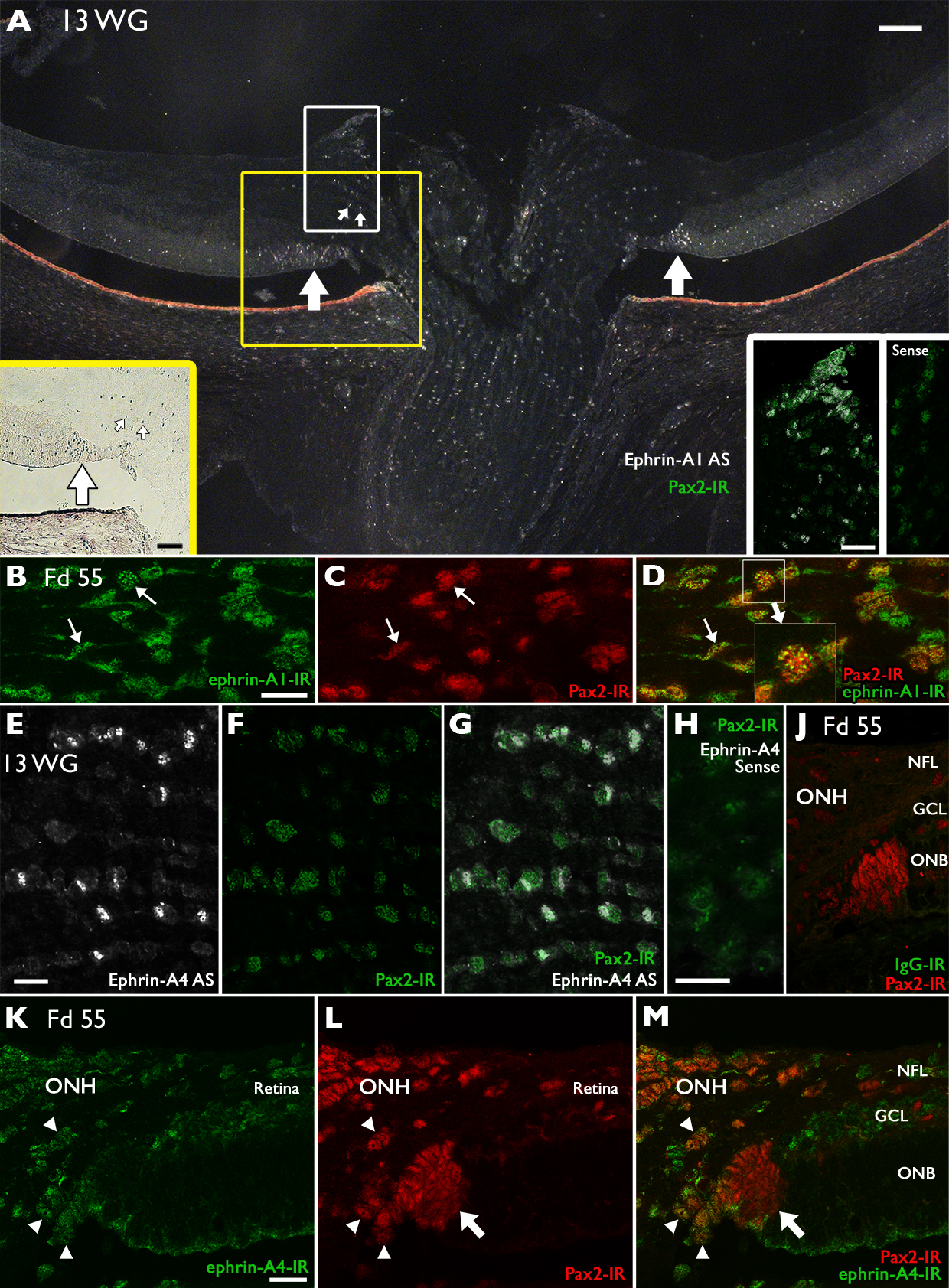
 Figure 6 of
Kozulin, Mol Vis 2009; 15:2649-2662.
Figure 6 of
Kozulin, Mol Vis 2009; 15:2649-2662.