Figure 4. In situ hybridization for Eph-A6
expression in a Fd 115 macaque retina. A: At the developing
fovea and in adjacent temporal retina, peak Eph-A6 mRNA
expression is detected in the inner ganglion cell layer (GCL) within
the foveal avascular area (bracket). Large arrows mark vessels at the
inner margin of the perifoveal plexus. The area between the two large
arrows is devoid of vessels. Smaller arrows indicate vessel profiles
deep in the GCL (GCL plexus), which is a characteristic of macular
vessels. B: Approximately 500 µm further into temporal retina,
vessels (arrows) are present in the inner and outer GCL, and levels of Eph-A6
mRNA in the GCL are lower, and more uniform across the depth of the
GCL, compared with A. Together, the images show two gradients
of Eph-A6 expression: a high-to-low gradient from inner to
outer GCL at the developing fovea (in A), and a high-to-low
gradient from fovea to periphery (top left GCL in A to lower
right GCL in B). Both A and B are at the same
magnification. Abbreviations: inner nuclear layer (INL); outer nuclear
layer (ONL).
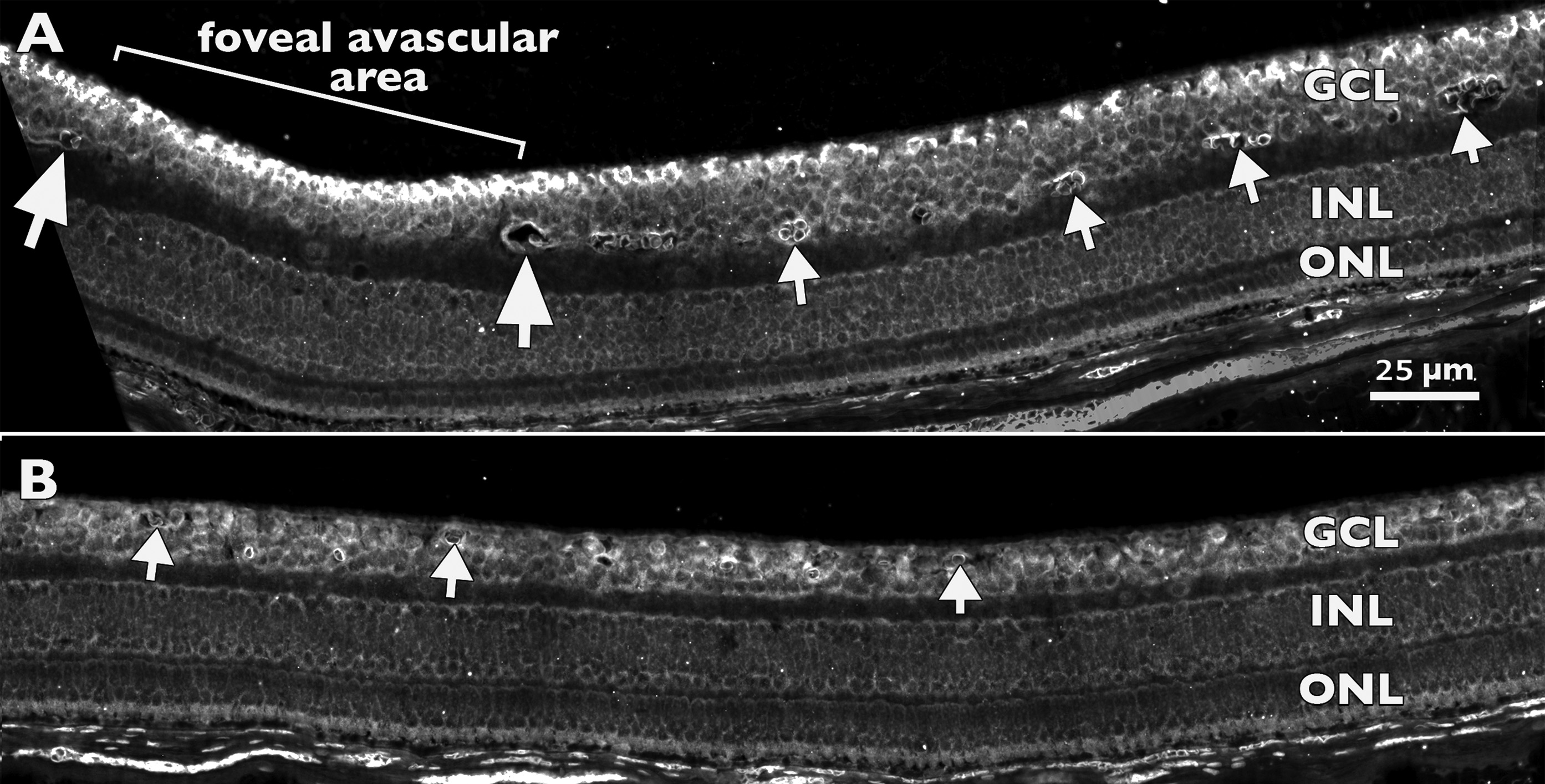
 Figure 4 of Kozulin, Mol Vis 2009; 15:2649-2662.