Figure 3. Optical densitometry profiles of
Eph-A6 mRNA expression. A-C: At Fd 55, Eph-A6
is expressed in all cell layers at each location, but at higher levels
in the outer nuclear layer (ONL) compared to other layers. D-F:
By Fd 80, Eph-A6 mRNA is higher in the ganglion cell layer
(GCL) than other layers. The mRNA is also higher at the fovea and rim
locations compared to nasal (F). At the fovea (D) and rim
(E) locations there is a gradient of Eph-A6 mRNA in the
GCL, with significantly higher levels of mRNA labeling present in the
inner GCL, and lower levels in the outer GCL (*p<0.0001,
Mann–Whitney test). G-J: A similar gradient of mRNA
expression was detected in the fovea and rim locations in a Fd 110
retina. K-L: Eph-A6 mRNA expression remains
relatively high in the GCL at Fd 145 at the fovea (K) and on the
rim (L), where an inner-to-outer gradient of expression in the
GCL is still detected. Eph-A6 expression is relatively lower
and uniform nasal to the fovea (M). Abbreviations: inner nuclear
layer (INL); inner plexiform layer (IPL).
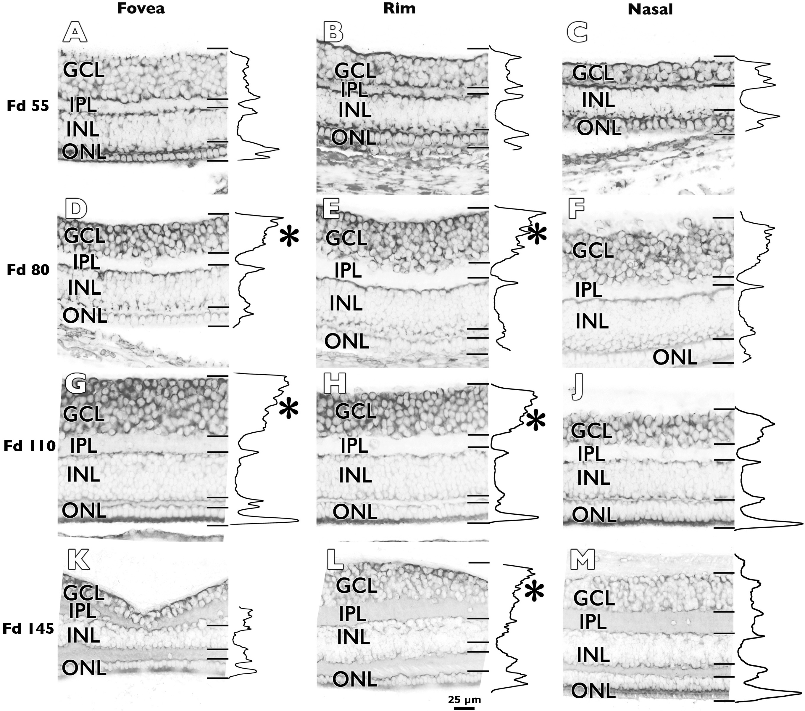
 Figure 3 of Kozulin, Mol Vis 2009; 15:2649-2662.