Figure 7. Apoptosis Inducing Factor localization in triamcinolone acetonide treated BRECs. Apoptosis inducing factor (AIF) immuno-labeling
was performed in control cells treated with the TA vehicle (1% ethanol; A-C). A cytoplasmic labeling is seen. This is also the case in cells treated with 0.1 mg/ml TA for 72h (D-F). In cells treated with TA for 5 days (G-I), cells with condensed nuclei (arrows in H and I) present a nuclear staining for AIF. Nuclei were stained with DAPI (A, D, G). Scale bar represents 50 µm.
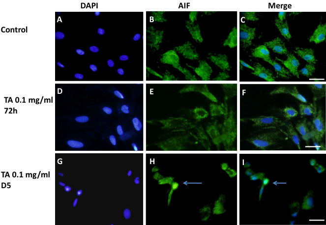
 Figure 7 of
Valamanesh, Mol Vis 2009; 15:2634-2648.
Figure 7 of
Valamanesh, Mol Vis 2009; 15:2634-2648.