Figure 7. Effects of BOL-303242-X and
dexamethasone (DEX) on p65 NFκB phosphorylation and IκB-α protein
levels in human corneal epithelial cells. Cells were pretreated with
vehicle, BOL-303242-X, or DEX for 2 h, and then further treated with
vehicle, IL1ß, IL-1ß plus BOL-303242-X, or DEX in basic EpiLife medium
for 10 min. Cell lysates were prepared and resolved by SDS-PAGE.
Western blotting was performed with phospho-p65 NFκB followed by p65
NFκB antibodies, or with IκB-α followed by keratin antibodies. The top
images show representative blots for phosphorylated p65 NFκB (pp65
NFκB), total p65 NFκB, IκB-α, and keratin. At the bottom, the results
of the densitometric quantification of the blots for pp65NFκB (left
panel) and IκB-α (right panel) phosphorylated:total p65 NFκB or
IκB-α:keratin ratios are shown. The data are expressed as % of IL-β.
Data are means±SEM from three to four independent experiments. The
asterisk indicates a p≤0.05 versus IL-1ß. One-way ANOVA followed by the
Dunnett’s test on raw data for pNFκB and IκB-α. In the figure, BOL
represents BOL-303242-X.
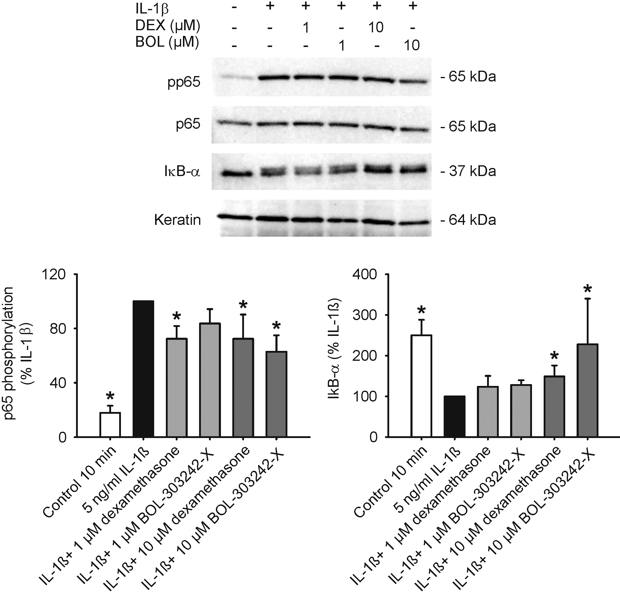
 Figure 7 of Zhang, Mol Vis 2009; 15:2606-2616.