Figure 9. Ocular hypertension induces
aberrant expression of phosphorylated neurofilament heavy chain.
Fluorescence photomicrographs show representative middle regions of
right eye (control; A, B) and left eye (lasered; C,
D) mouse retinas, with 10% hydroxystilbamidine methanesulfonate
(OHSt) applied to the superior colliculi for 1 week, that were
processed for RT97 immunofluorescence 17 days after laser
photocoagulation. Micrographs were taken under ultraviolet and
fluorescein filters to examine OHSt or RT97 immunofluorescence,
respectively. A, B: In the control (right-eye) retina,
OHSt-labeled RGCs show typical distribution (A) and RT97
immunofluorescence is restricted to axonal bundles (B). C,
D: In the lasered (left-eye) retina, the distribution of retinal
ganglion cells (RGCs) retrograde-labeled with OHSt is restricted to the
lower part of the picture (C), which under the fluorescein
filter (D) shows a lack of RT97 staining within OHSt+
RGCs. Abnormal phosphorylated neurofilament heavy chain expression,
which depicts several intensely RT97-stained RGCs and RT97 beaded
axons, is concentrated in the region that lacks OHSt backlabeled RGCs.
Scale bars for A-D=50 µm.
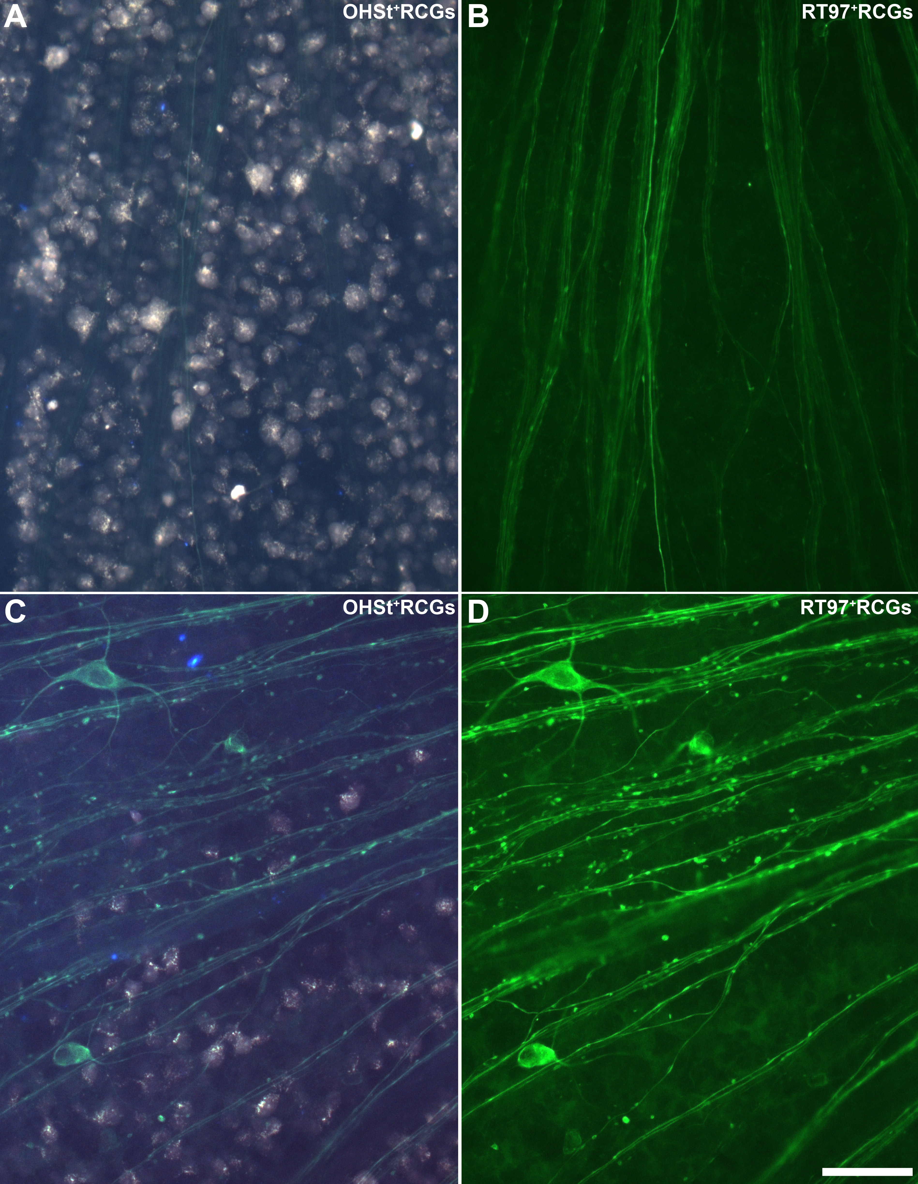
 Figure 9 of Salinas-Navarro, Mol Vis 2009; 15:2578-2598.