Figure 4. Histograms illustrating evolving
changes in the electroretinograms. Average values (mean±SEM) of the
positive scotopic threshold response (pSTR) and a- and b-waves are
presented as percentages of their basal values in three different
experimental groups that were analyzed at increasing survival intervals
and sacrificed at 17 (A), 35 (B), or 63 (D) days
after lasering. At 24 h after laser treatment, a large reduction can be
seen in the amplitudes of all the registered waves when compared to
their basal levels. These reductions are maintained over time and do
not recover significantly even for the animal group examined at 8 weeks
after lasering (C), suggesting that laser treatment induces a
permanent functional impairment within the innermost, inner and outer
retina.
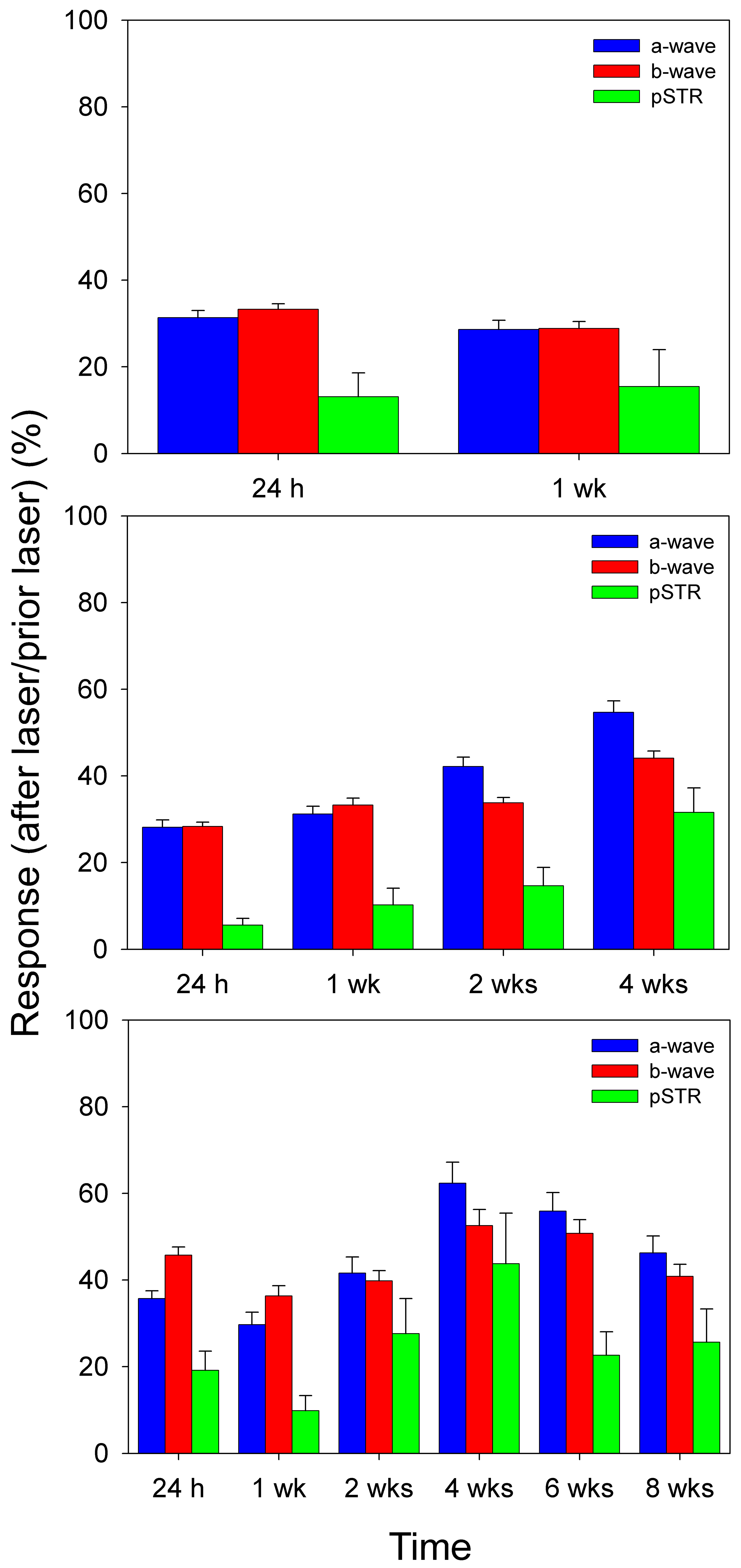
 Figure 4 of Salinas-Navarro, Mol Vis 2009; 15:2578-2598.