Figure 10. Different geographical
distribution of RT97-labeled and OHSt-labeled retinal ganglion cells in
retinas with ocular hypertension. These examples of representative left
retinas in experimental animals 17 days after lasering the left eye
show retinal ganglion cells (RGCs) labeled with 10% hydroxystilbamidine
methanesulfonate (OHSt) and RT97, illustrating the near confinement of
abnormal expression of RT97 to sectors of the retina that lack OHSt
backlabeled RGCs; meanwhile, sectors containing OHSt+ RGCs
present fewer RGCs with abnormal RT97 staining. To identify RGCs
capable of retrograde axonal transport, OHSt was applied to both
superior colliculi 1 week before sacrifice. Retinal whole-mounts were
immunostained with RT97 antibodies to identify RT97+ RGCs.
OHSt+ RGC isodensity maps were generated by assigning a
color code to each of the 36 subdivisions of each individual frame
according to its density value within a 45-step color scale, ranging
from 0 (dark blue) to 5,625 RGCs/mm2 or higher (red). RT97+
RGCs are represented as dots over the outline of the retinal
whole-mount. A, B: A whole-mount of a representative
experimental left retina doubly labeled with OHSt (A) and RT97 (B)
illustrates the typical topological distribution of 12,821 OHSt+
RGCs and 415 RT97+ RGCs throughout the retina in these
experiments. Note the lack of correspondence between areas containing
OHSt-labeled RGCs (A), which are restricted to a large wedge
located between the 5 and 10 o’clock positions, and the RT97+
RGCs (B), which are distributed mainly in the opposite area of
the retina. The dorsal pole of the retina is orientated at 12 o’clock. C,
D: Another whole-mounted representative experimental left retina
doubly labeled with OHSt (C) and RT97 (D) illustrates the
topological distribution of 10,911 OHSt+ RGCs and 269 RT97+
RGCs throughout the retina in these experiments. Note the lack of
correspondence between areas containing OHSt-labeled RGCs (C)
which are restricted to a large wedge between 4:30 and 8:30 o’clock,
and the RT97+ RGCs (D) found mainly in the opposite
area of the retina. The dorsal pole of the retina is orientated at 12
o’clock (scale bar=1 mm).
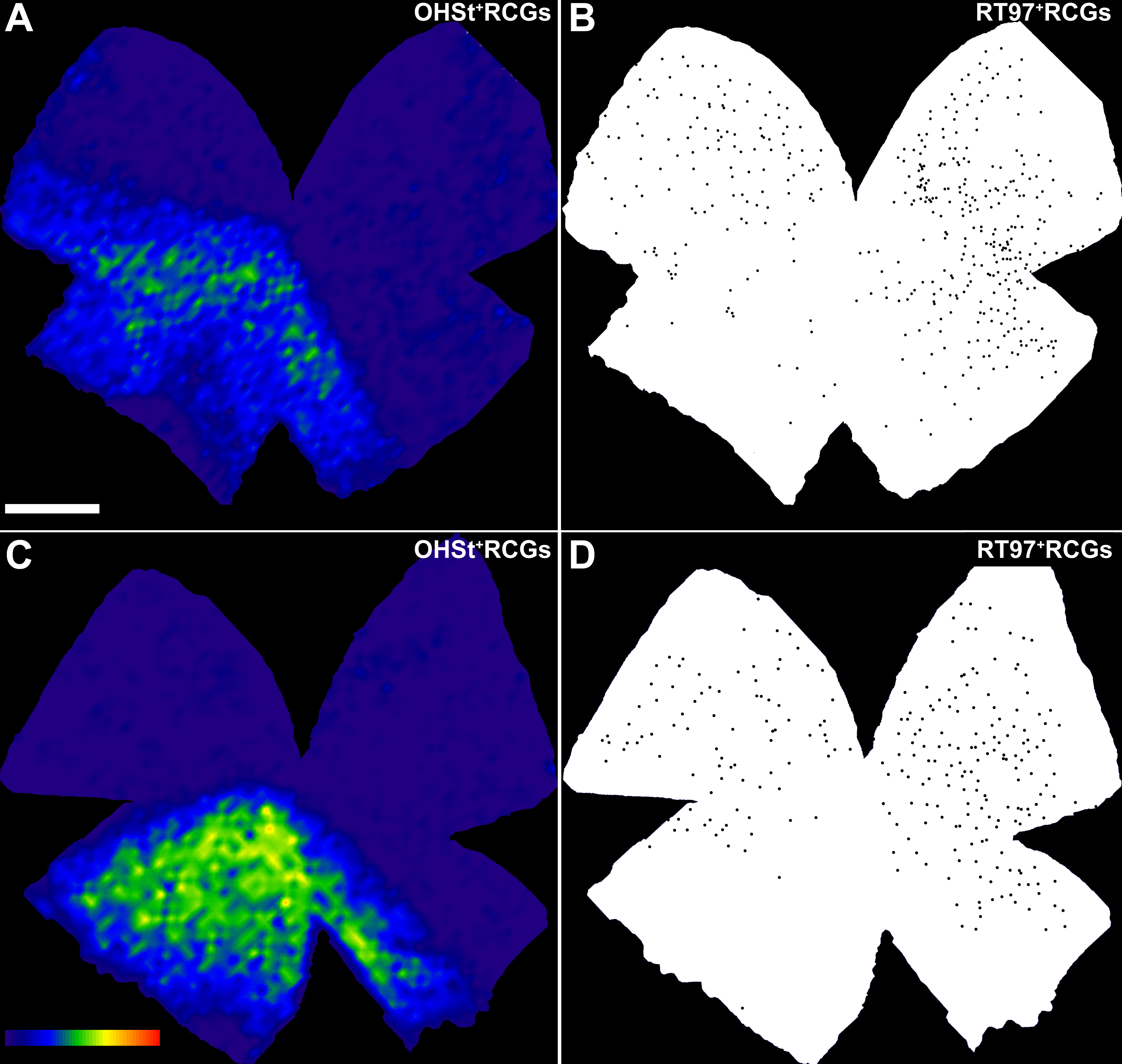
 Figure 10 of Salinas-Navarro, Mol Vis 2009; 15:2578-2598.