Figure 4. Western blots of proteins from human corneal stromal fibroblasts. HCS fibroblasts were grown to confluence in DMEM containing
5% FBS. The cell membranes were prepared and lysed in RIPA. The aliquots of proteins were then analyzed by western blotting.
The primary antibodies detected proteins of the correct molecular weight; Row 1, Magic Mark molecular weight standards; Row
2, empty; Row 3, positive control protein as detected protein as detected in HL60, HEK or HUVEC cells; Row 4, proteins as
detected in HCS fibroblasts. Primary antibodies include A: NOX1, B: NOX4, C: NOX5, D: p22 phox, E: p47 phox, F: p40 phox, G: p67 phox, and H: Rac.
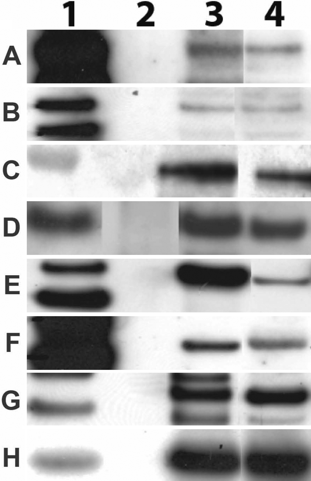
 Figure 4 of
O’Brien, Mol Vis 2009; 15:2535-2543.
Figure 4 of
O’Brien, Mol Vis 2009; 15:2535-2543.