Figure 2. Clinical photographs of affected
family members with LCD I. A and B: Slit lamp
appearance of a cornea of a proband at 16 years of age shows distinct
refractile lattice lines and diffuse opacification in the subepithelial
and anterior stromal layer in the left eye. C: The photograph
demonstrates central anterior stromal clouding in the proband’s right
eye. D: The photograph shows lattice opacities with the
appearance of new vessels in the subepithelial and anterior stromal
cornea of the proband’s mother, with recurrent disease in the right
eye. E: The image of the proband’s mother reveals thick linear
opacities and diffuse grayish-white clouding, which partly covers the
original lattice lines within the central area of the cornea in the
left eye.
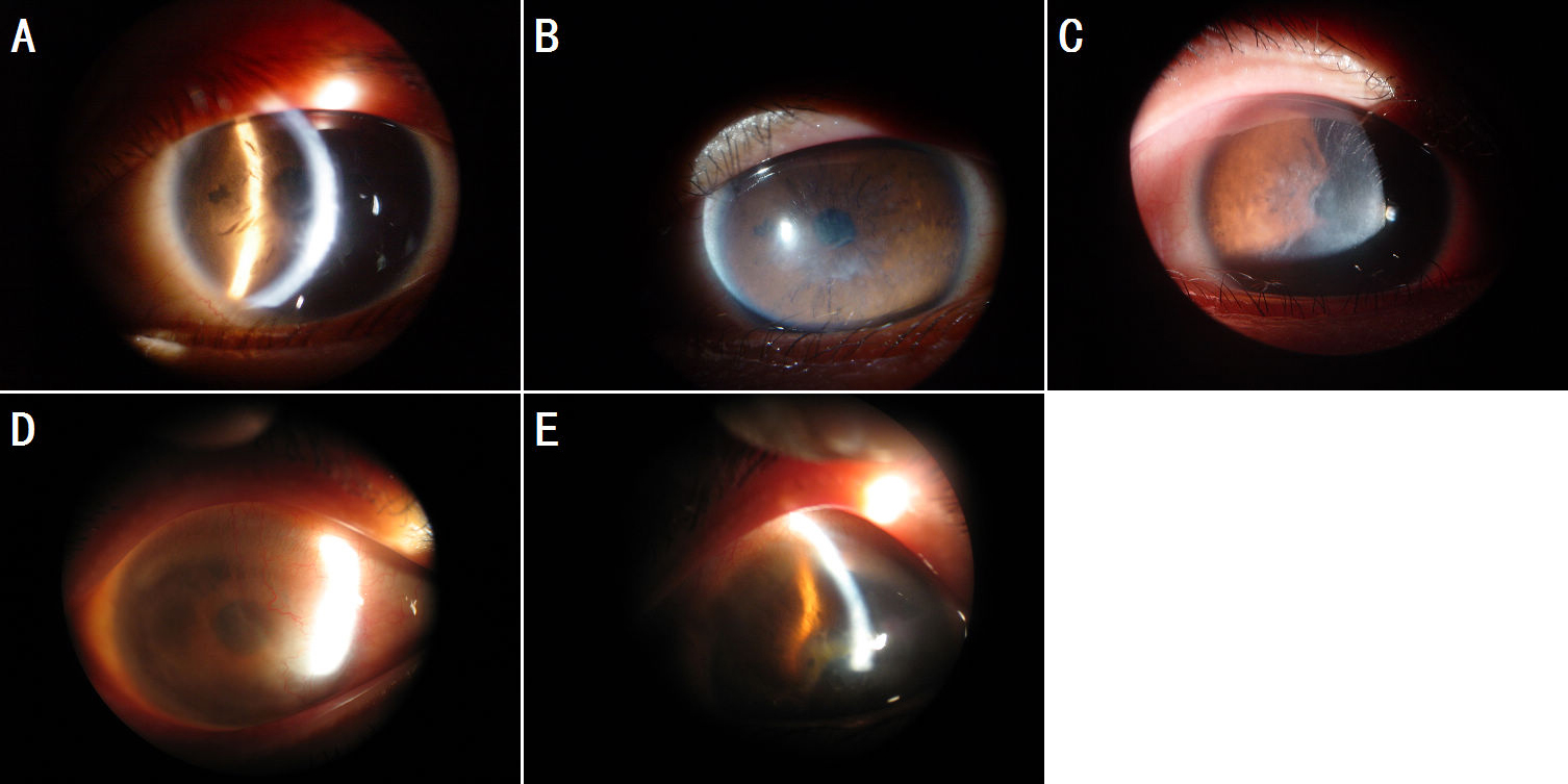
 Figure 2 of Zhang, Mol Vis 2009; 15:2498-2502.