Figure 2. Fundus photography. Images are right and left eye color fundus photograph composites of patient NH13424 performed using TOPCON
retinal camera at 2.45x magnification at 35 degrees. Composite images were created using Adobe Photoshop. Image cropping and
reflections are observed secondary to poor pupil dilatation. The images show significant and abnormal peripapillary hyperpigmentation
with fine granular pigmentation at the level of the retinal pigment epithelium (RPE).
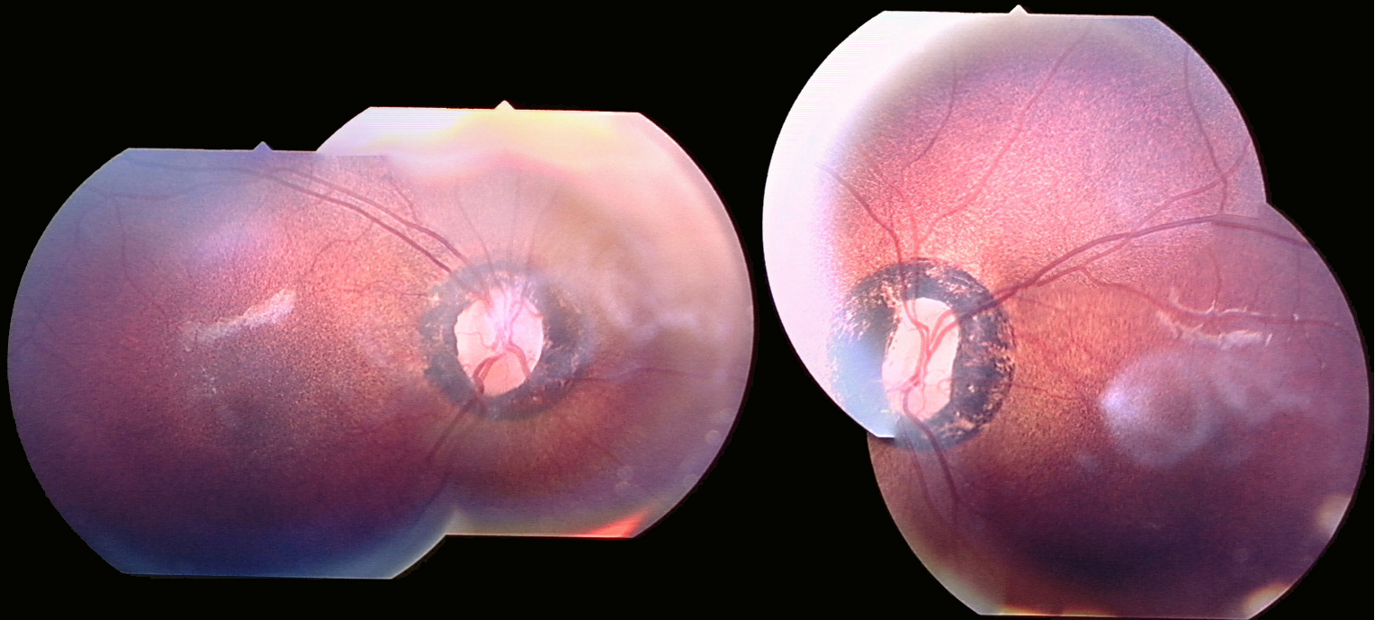
 Figure 2 of
Henderson, Mol Vis 2009; 15:2442-2447.
Figure 2 of
Henderson, Mol Vis 2009; 15:2442-2447.