Figure 2. SEM image of the outer region of human and porcine sclera. Original magnification was 5,000X in panel A and 20,000X in panel B. Scale bar=10 µm in both cases. From the pictures at lower magnification (A) it is possible to observe branching and anastomosis of the collagen bundles to form dense connective tissue. The bundles
were of varying thickness and width and often intertwined with each other. Porcine bundles looked thicker at lower magnification,
but still showed a similar arrangement. Moreover, higher magnification (B) did not show any difference between human and porcine sclera in the diameter of the single collagen fibers
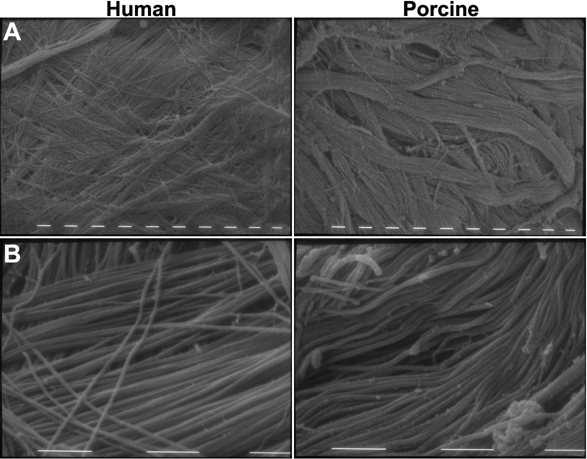
 Figure 2 of
Nicoli, Mol Vis 2009; 15:259-266.
Figure 2 of
Nicoli, Mol Vis 2009; 15:259-266.