Figure 1. Histological microscopic sections of human and porcine sclera stained with hematoxylin-eosin. Empty lacunae between fibers
are an artifact due to tissue preparation. Original magnification was 10X in panel A and 4X in panel B. In both human and porcine scleras, scattered small fibrocyte nuclei are dispersed between the bundles of interwoven collagen
fibers. In porcine sclera, thicker and more disorganized collagen bundles are visible. From the 4X magnification (B) it is possible to appreciate differences in thickness between human and porcine sclera.
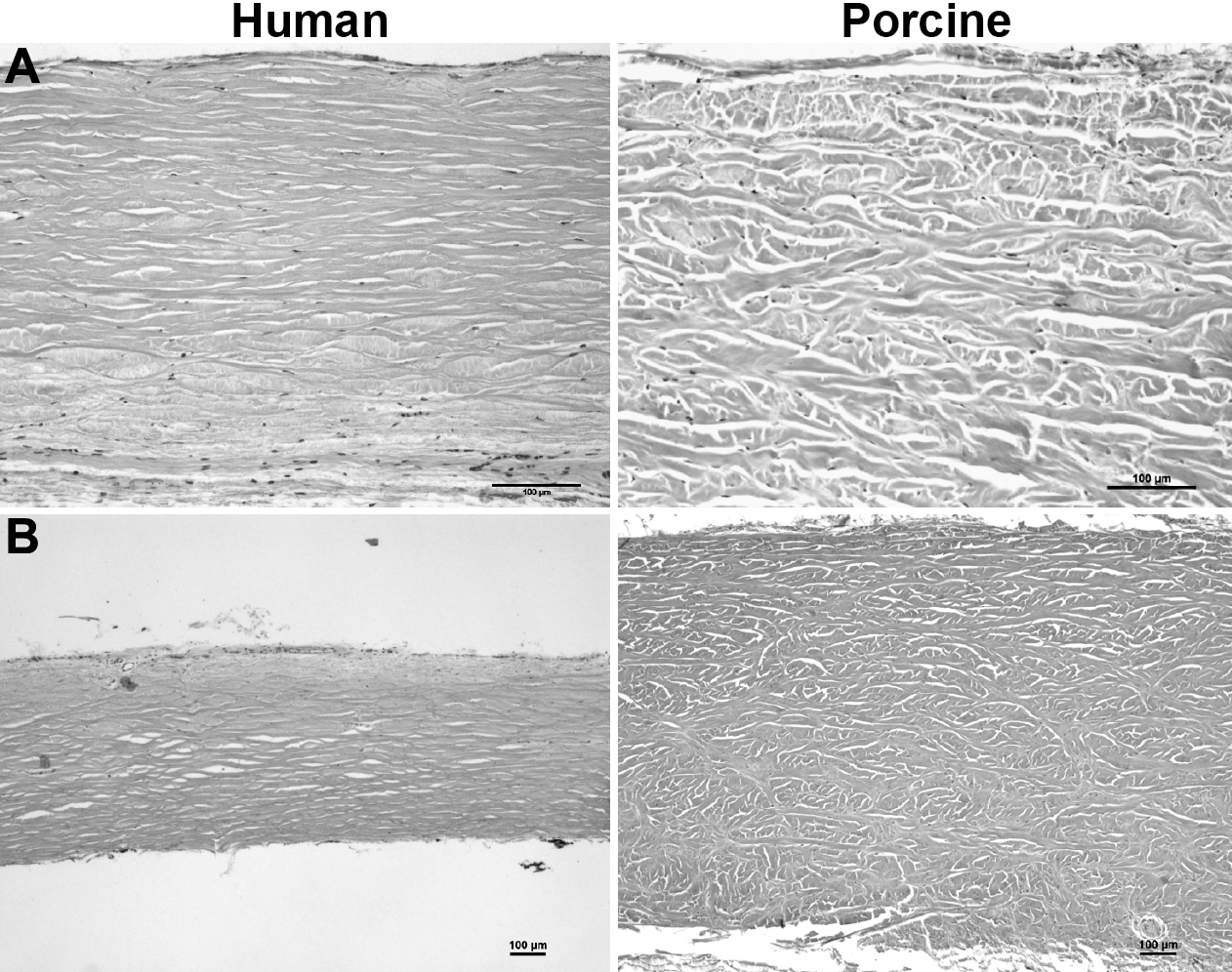
 Figure 1 of
Nicoli, Mol Vis 2009; 15:259-266.
Figure 1 of
Nicoli, Mol Vis 2009; 15:259-266.