Figure 5. Immunofluorescence indicates
that SAT1 expression is associated with the leading edge and body of
pterygium. SAT1 immunofluorescence (IF) is shown in red and nuclei are
DAPI labeled in blue. CE, corneal epithelium; Pt, pterygium; Cj,
conjunctiva. A: SAT1 is detectable throughout the body of
the pterygium with a strongly labeled clump of cells at the leading
edge. B: Magnified view is presented of the clump of cells
strongly positive for SAT1 at the apex of the pterygium body. C:
No SAT1 IF is apparent in limbus and normal cornea from the other side
of the same eye (section torn in corneal stroma).
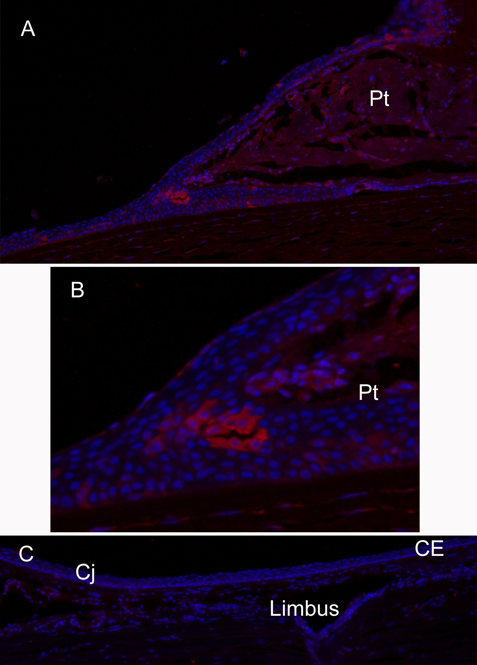
 Figure 5 of Jaworski, Mol Vis 2009; 15:2421-2434.