Figure 3 of
Jaworski, Mol Vis 2009; 15:2421-2434.
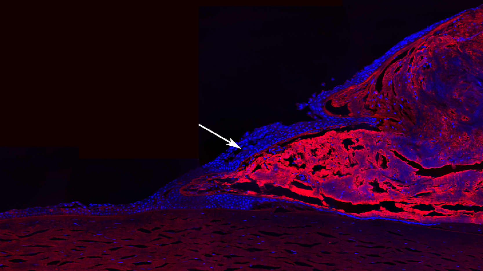
Figure 3.
The abundance of clusterin in pterygium is illustrated by immunofluorecence. Labeling for clusterin is in red, nuclei are blue (DAPI). The arrow indicates cells enveloping the body of the pterygium that are clusterin negative.
 Figure 3 of Jaworski, Mol Vis 2009; 15:2421-2434.