Figure 2. Immunofluorescence shows the
localization of keratin 12 and 13 in normal ocular surface and
pterygium. Krt12 is labeled in red, Krt13 in green, and nuclei are DAPI
labeled in blue. A: Krt12 and Krt13 are segregated in corneal
epithelium and conjunctival epithelium with a boundary at the limbus:
Cj, conjunctiva; Lm, limbus; Ce, corneal epithelium. B: DAPI
staining of (A) indicates the nuclei of conjunctiva, limbal
region, and cornea. C: Corneal epithelium Ce, is Krt12
positive. The leading edge of a pterygium, Le, has mixed Krt12-
and Krt13-positive cells. D: DAPI stain of (C) shows the
nuclei of the leading edge region and cornea.
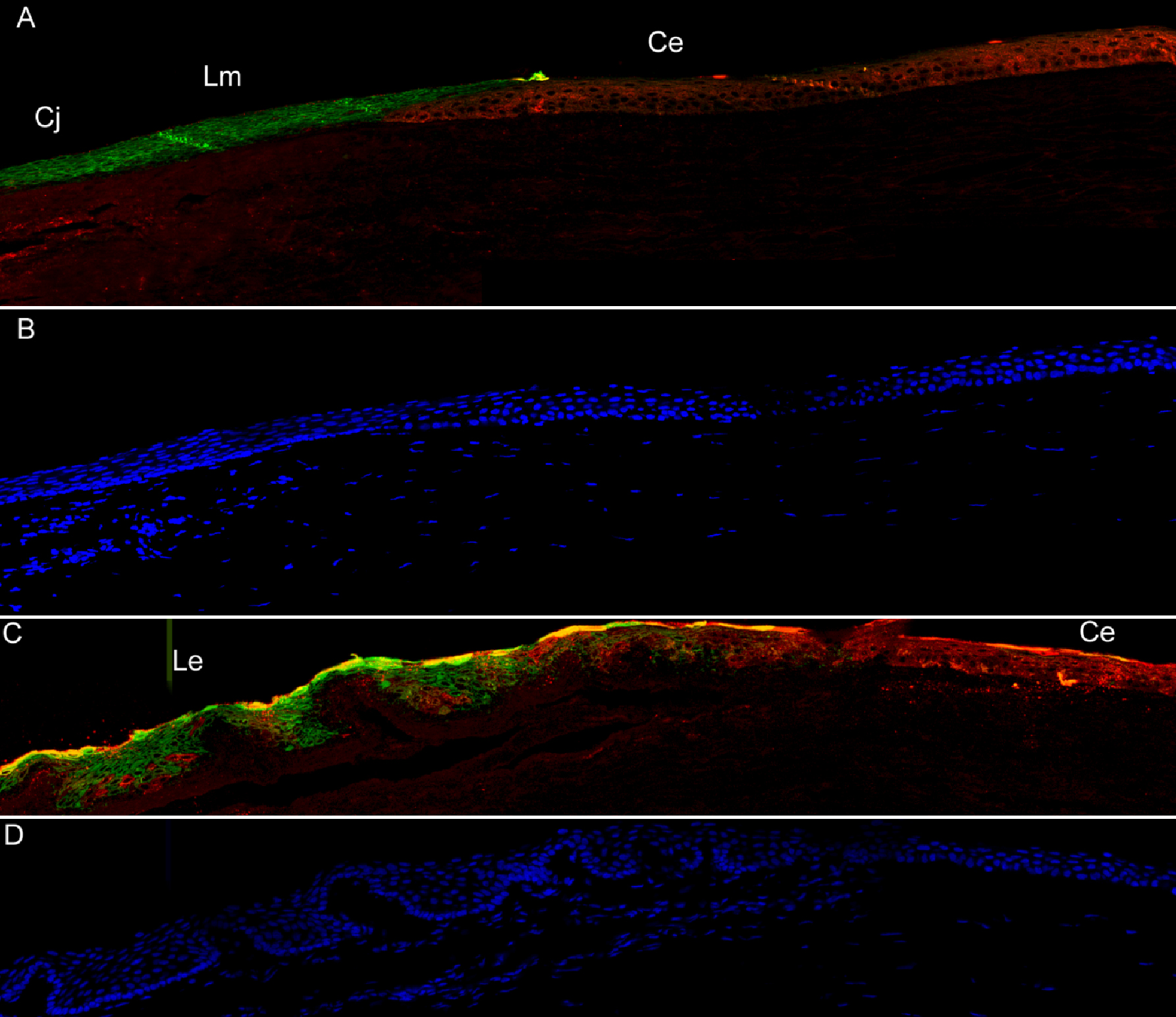
 Figure 2 of Jaworski, Mol Vis 2009; 15:2421-2434.