Figure 1 of
Ghahghaei, Mol Vis 2009; 15:2411-2420.
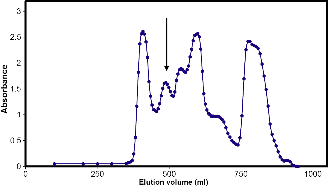
Figure 1.
Fractionation of dogfish lens extract on a 3×150 cm Sephacryl S300 column. The position of the α-crystallin peak (490 ml) is indicated with an arrow. The central 50% of the peak was rechromatographed under the same conditions.
 Figure 1 of Ghahghaei, Mol Vis 2009; 15:2411-2420.