Figure 4. Western blot analysis of the distribution of ferritin H chains within the layers of fiber cells (L1-L6) from noncataractous
and cataractous lenses. Fiber cell homogenates (50 µg protein/sample) were separated by 15% SDS-PAGE under reducing conditions.
Anti-canine ferritin H-chain antibodies were used to identify ferritin H chains. Canine heart ferritin was used as a standard.
The representative blots show the H-chain distribution in lens fiber cells from a 1-year-old dog’s noncataractous lens and
a 10-year-old dog’s lens with an early cataract.
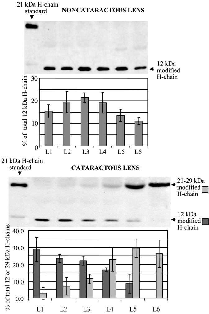
 Figure 4 of
Goralska, Mol Vis 2009; 15:2404-2410.
Figure 4 of
Goralska, Mol Vis 2009; 15:2404-2410.