Figure 2. Total protein profile of different layers of fiber cells. A 50 μg sample (from a 10-year-old dog’s transparent lens) of protein
homogenate from each layer was separated by 15% SDS PAGE under reducing conditions. Proteins were stained with coomassie blue.
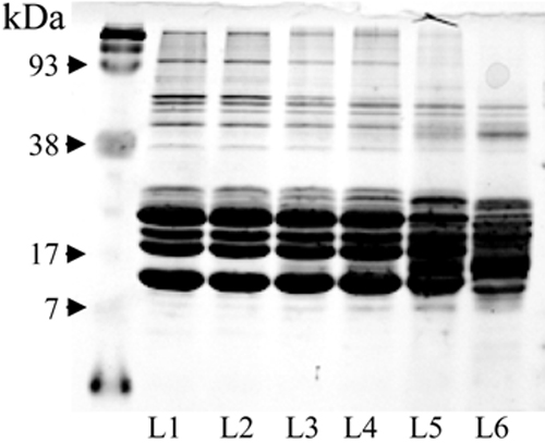
 Figure 2 of
Goralska, Mol Vis 2009; 15:2404-2410.
Figure 2 of
Goralska, Mol Vis 2009; 15:2404-2410.