Figure 7. Confocal double-label
immunocytochemical localization of Mel1a and ZO-1 in Xenopus
corneal whole mounts. A: Mel1a and ZO-1 immunolabeling is
observed on the lateral plasma membrane, with some immunoreactivity
also occurring in the cytoplasm. Mel1a (red) labeling is present
predominantly as very broad bands on the lateral membranes, including
the obliquely-oriented lateral membranes (arrows). The plasma membrane
has an abundance of yellow labeling, indicative of a very close
proximity of red Mel1a and green ZO-1. The ZO-1 labeling was not as
broadly distributed on the lateral membrane. The inset illustrates the
72° rotation on the x-axis of the image in A, indicating the
orientation relative to the viewer’s eye in B. B:
Three-dimensional reconstructions of confocal z-stacks of optical
slices were rotated at 72° degrees on the x-axis to enable optimal
viewing of the pattern of immunolabeling. Arrows are provided as
reference points to indicate the same points on panel A. The
rotated image shows that the green ZO-1 labeling is generally located
apically to the red Mel1a labeling. The ZO-1 labeling appears as a
relatively narrow continuous band of green labeling on the lateral
plasma membrane, whereas a much broader band of red Mel1a labeling
appears directly basal to the ZO-1 label. Significant yellow labeling
is observed, indicating that the Mel1a receptor is in very close
proximity to ZO-1. The confocal images in both panels are comprised of
seven optical slices of 400 nm each in the z-series. The magnification
bar (B) represents 20 µm.
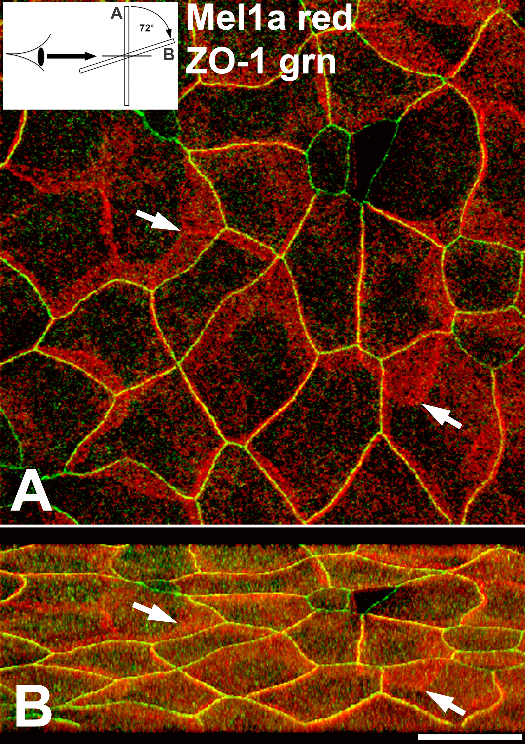
 Figure 7 of Wiechmann, Mol Vis 2009; 15:2384-2403.