Figure 6. Localization of Mel1a and Mel1b
in progressive confocal optical slices of Xenopus corneal
epithelium. A: Image of the most superficial surface of the
surface corneal epithelium. Note the predominance of yellow (merged red
and green) labeling of most lateral membranes, with a lesser amount of
interdigitated red Mel1a labeling (note arrowheads indicating an
example of this). B-F: As the 0.4-µm slices progress deeper
into the corneal epithelium layer , there is not a transition from red
to green labeling as was seen with Mel1a-Mel1c, but instead the
predominance of yellow labeling with some interspersed red labeling is
maintained throughout all slices. Nuclei are stained with DAPI. The
magnification bar (F) represents 20 µm.
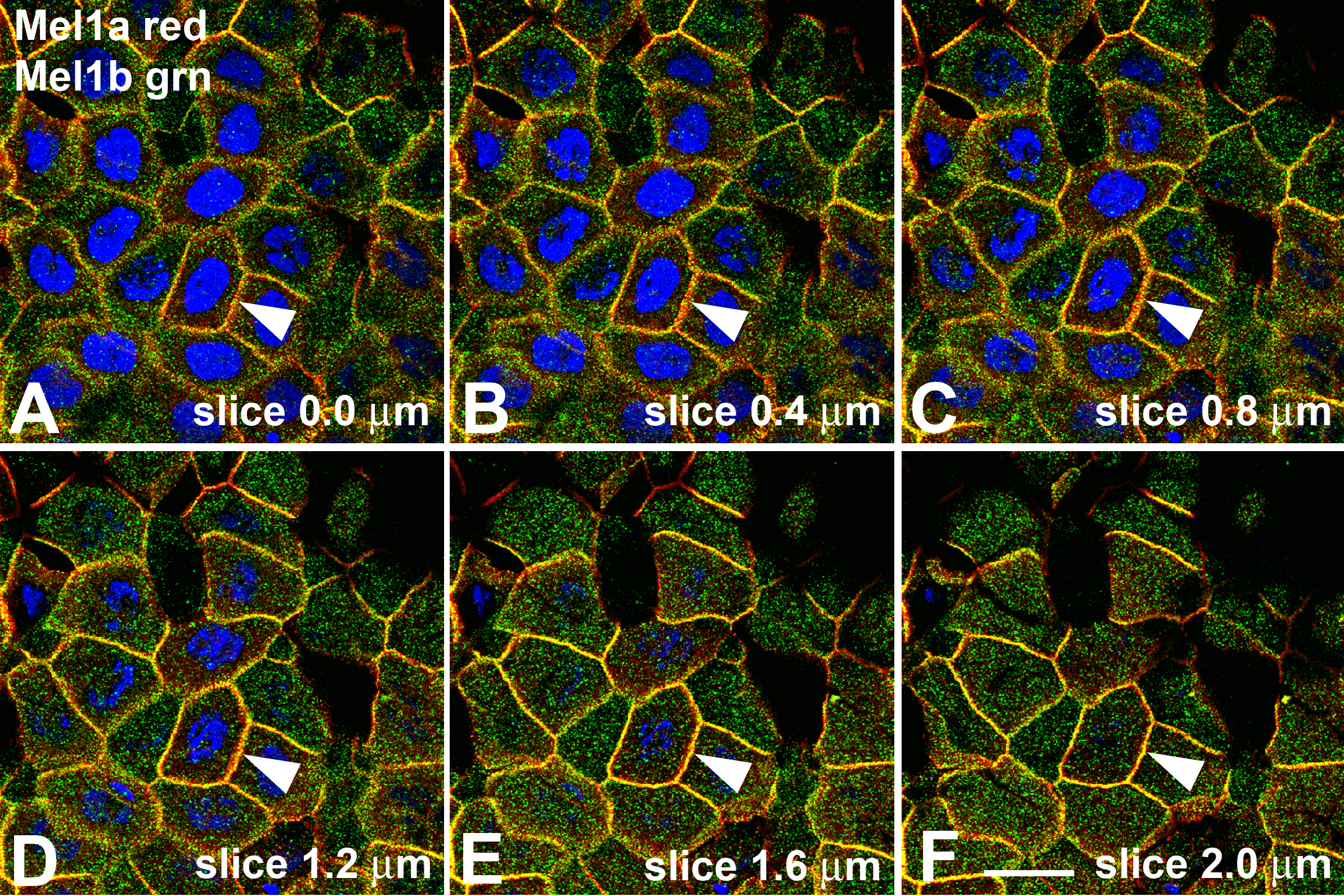
 Figure 6 of Wiechmann, Mol Vis 2009; 15:2384-2403.