Figure 4. Localization of Mel1a and Mel1c
in progressive confocal optical slices of Xenopus corneal
epithelium. A: Image of the most superficial surface of the
surface corneal epithelium. Note that only the red Mel1a
immunoreactivity is present on the lateral membranes. B-F: As
the 0.4 µm slices progress deeper into the corneal epithelium layer,
the Mel1a immunoreactivity lessens, whereas the green Mel1c
immunoreactivity increases (note arrowheads indicating an example of
this), indicating that the Mel1c receptor is located basal to the Mel1a
receptor. Nuclei are stained with DAPI. The magnification bar (F)
represents 20 µm.
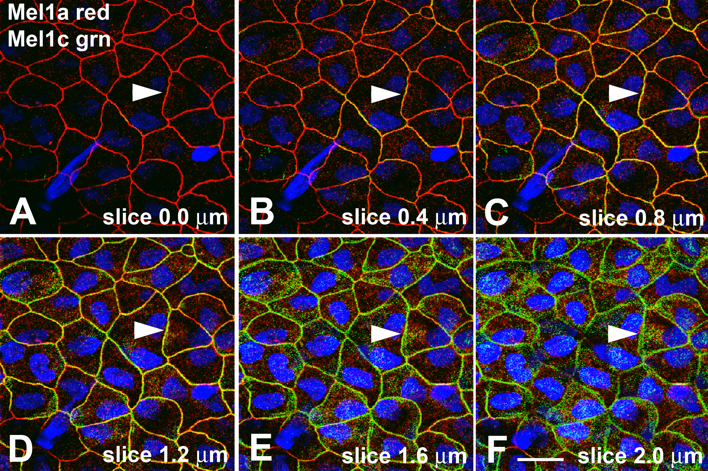
 Figure 4 of Wiechmann, Mol Vis 2009; 15:2384-2403.