Figure 3. Confocal double-label
immunocytochemical localization of Mel1a and Mel1c in Xenopus
corneal whole mounts. A: The specimen shown was obtained in the
mid-light period (12N). Both Mel1a and Mel1c immunolabeling is observed
on the lateral plasma membrane, with some immunoreactivity also
occurring in the cytoplasm. The labeling of the plasma membrane
displays distinct areas of Mel1a (red), Mel1c (green), and both
receptors (yellow). Arrows are provided as reference points to indicate
the same points on B. The inset illustrates the 72° rotation on
the x-axis of the image in A, indicating the orientation
relative to the viewer’s eye in B. B: Three-dimensional
reconstructions of confocal z-stacks of optical slices were rotated at
72° degrees on the x-axis to enable optimal viewing of the pattern of
immunolabeling. The rotated image shows that the red Mel1a labeling is
generally located apically to the green Mel1c labeling. The Mel1a
labeling is seen as a relatively broad continuous band of red label on
the lateral plasma membrane of the majority of surface CE cells. A
somewhat broader band of green Mel1c labeling appears directly basal to
the Mel1a label. Some yellow labeling is occasionally observed,
indicating some co-localization of Mel1a and Mel1c. There are many
areas in the red Mel1a band in which yellow labeling is interspersed
between areas of red Mel1a labeling, suggesting that some green
Mel1c-labeled receptor is interdigitated among the Mel1a-labeled
receptor. The confocal images in both panels are comprised of 13
optical slices of 400 nm each in the z-series. The magnification bar (B)
represents 20 µm.
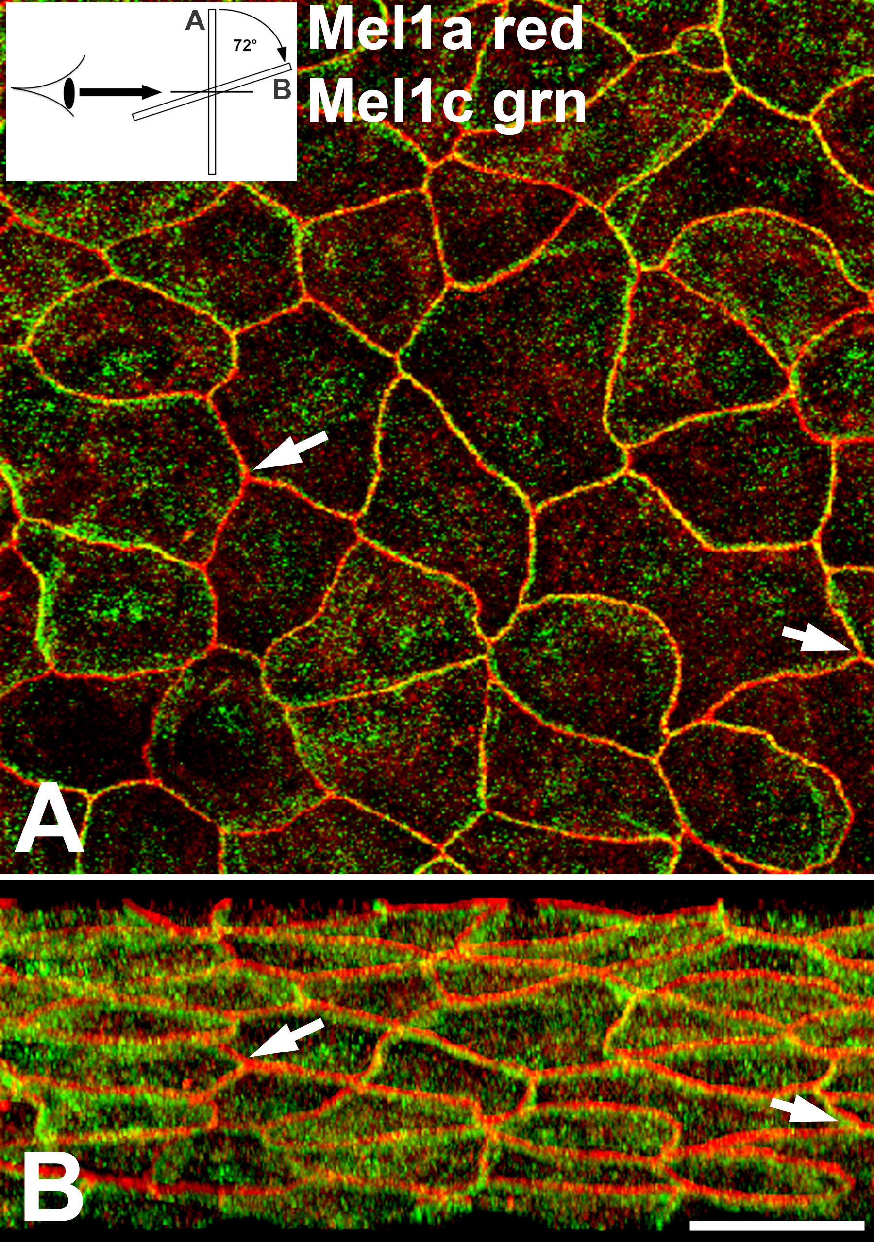
 Figure 3 of Wiechmann, Mol Vis 2009; 15:2384-2403.