Figure 2. Mel1a, Mel1b, and Mel1c
double-label immunocytochemistry of cryostat sections of Xenopus
laevis corneal epithelium. A and C: Corneas
obtained at 12:00 noon (12N) in the light. B and D:
Corneas obtained at 12:00 midnight (12M) in the dark. Sections were
immunolabeled with Mel1a and either Mel1b or Mel1c receptor antibodies.
Mel1a labeling is represented in red, and Mel1b and Mel1c labeling is
represented in green. Yellow indicates regions of co-localization of
the red and green signal. Melatonin receptors are expressed in the
surface epithelium, but their relative levels of expression and
distribution change between 12N and 12M. Arrows indicate the
immunolabeled plasma membranes of the surface epithelium. Nuclei are
stained with DAPI. The magnification bar (D) represents 20 µm.
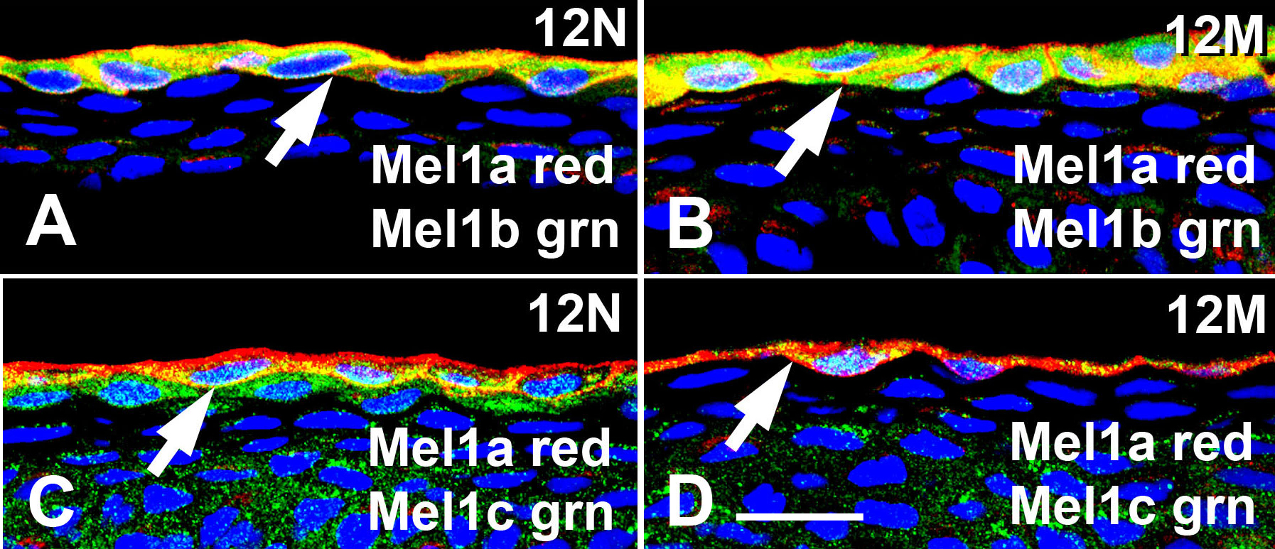
 Figure 2 of Wiechmann, Mol Vis 2009; 15:2384-2403.