Figure 1. Mel1a, Mel1b, and Mel1c
immunocytochemistry of cryostat sections and whole mounts of Xenopus
laevis corneal epithelium. A-C: Cryostat sections of
corneas obtained during the light period were immunolabeled with Mel1a,
Mel1b, or Mel1c receptor antibodies. Arrows in panel A indicate the
immunolabeled plasma membranes of the surface epithelium. D:
The control specimen was processed in the absence of primary antibody. E-G:
Whole mount preparations of corneas obtained during the light period
were immunolabeled with Mel1a, Mel1b, or Mel1c receptor antibodies. H:
The control whole mount specimen was processed in the absence of
primary antibody. Primary antibodies were labeled with secondary
antibody conjugated to AlexaFluor 488 (green fluorescence). Most of the
Mel1a receptor labeling (A and E) occurs in the lateral
plasma membrane of the surface epithelium, whereas there is a higher
proportion of Mel1b (B and F) and Mel1c (C and G)
labeling also present in cytoplasmic compartments in addition to the
lateral membranes. Note that no specific immunolabeling is detected in
the control specimens (D and H). Nuclei are stained with
DAPI. The magnification bar (H) represents 20 µm.
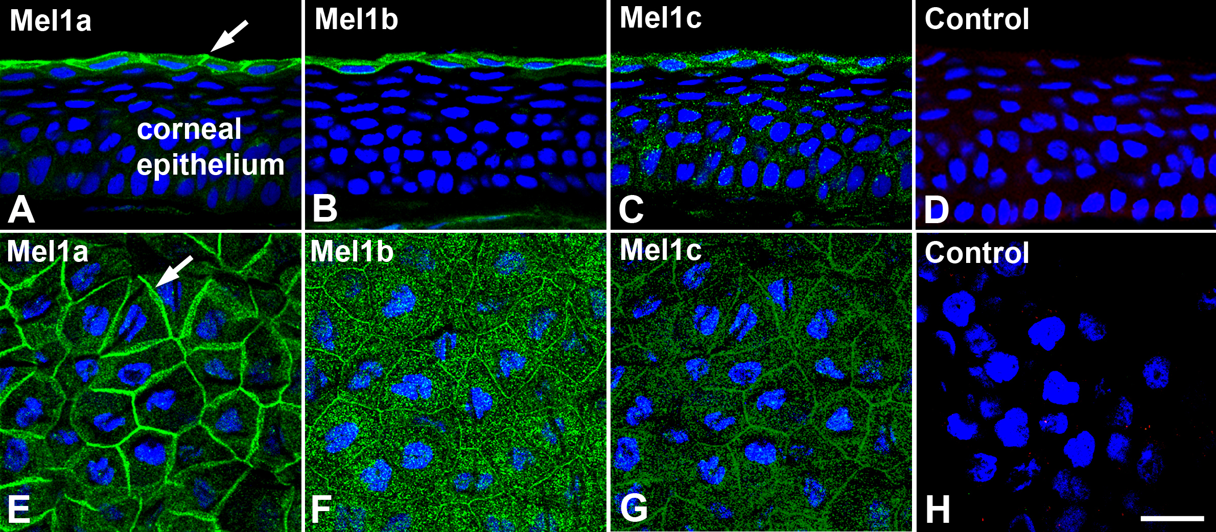
 Figure 1 of Wiechmann, Mol Vis 2009; 15:2384-2403.