Figure 7. Immunofluorescent staining of
MIF in corneal and conjunctival epithelia. Immunofluorescence staining
demonstrated the presence of MIF (red) in corneal epithelia (CE:
arrows) and conjunctival epithelia (CJE: arrow heads) at all time
points. The staining was detected in normal (A, E, I,
and M) and BTX-B injected mice (B, F, J,
and N) and saline-injected mice (D, H, L,
and P) and showed no difference (C, G, K,
and O). Negative control with no staining. Isotype control was
omitted with no staining. Scale bar: 50 μm.
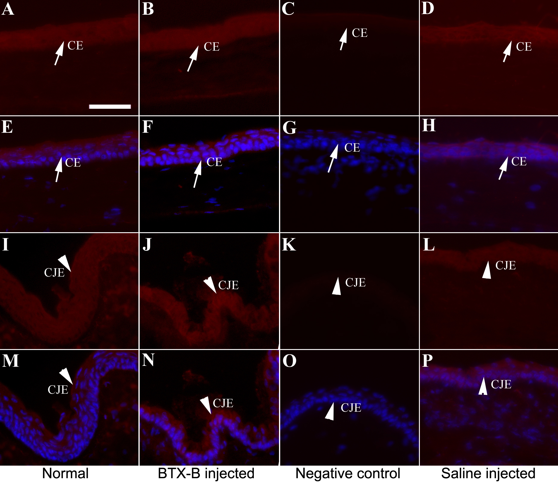
 Figure 7 of Zhu, Mol Vis 2009; 15:250-258.