Figure 6. Immunofluorescent staining with
IL-1β specific antibody in corneal and conjunctival epithelia.
Immunofluorescence staining with IL-1β specific antibody (red) in
corneal epithelia (CE: arrows) and conjunctival epithelia (CJE: arrow
heads) at all time points. The staining for IL-1β localized in CE and
CJE was strong in BTX-B injected mice. Nuclear staining in blue. (F-J)
and (P-T) merged pictures. Images of isotype and negative
control were omitted with no staining. Scale bar: 50 μm.
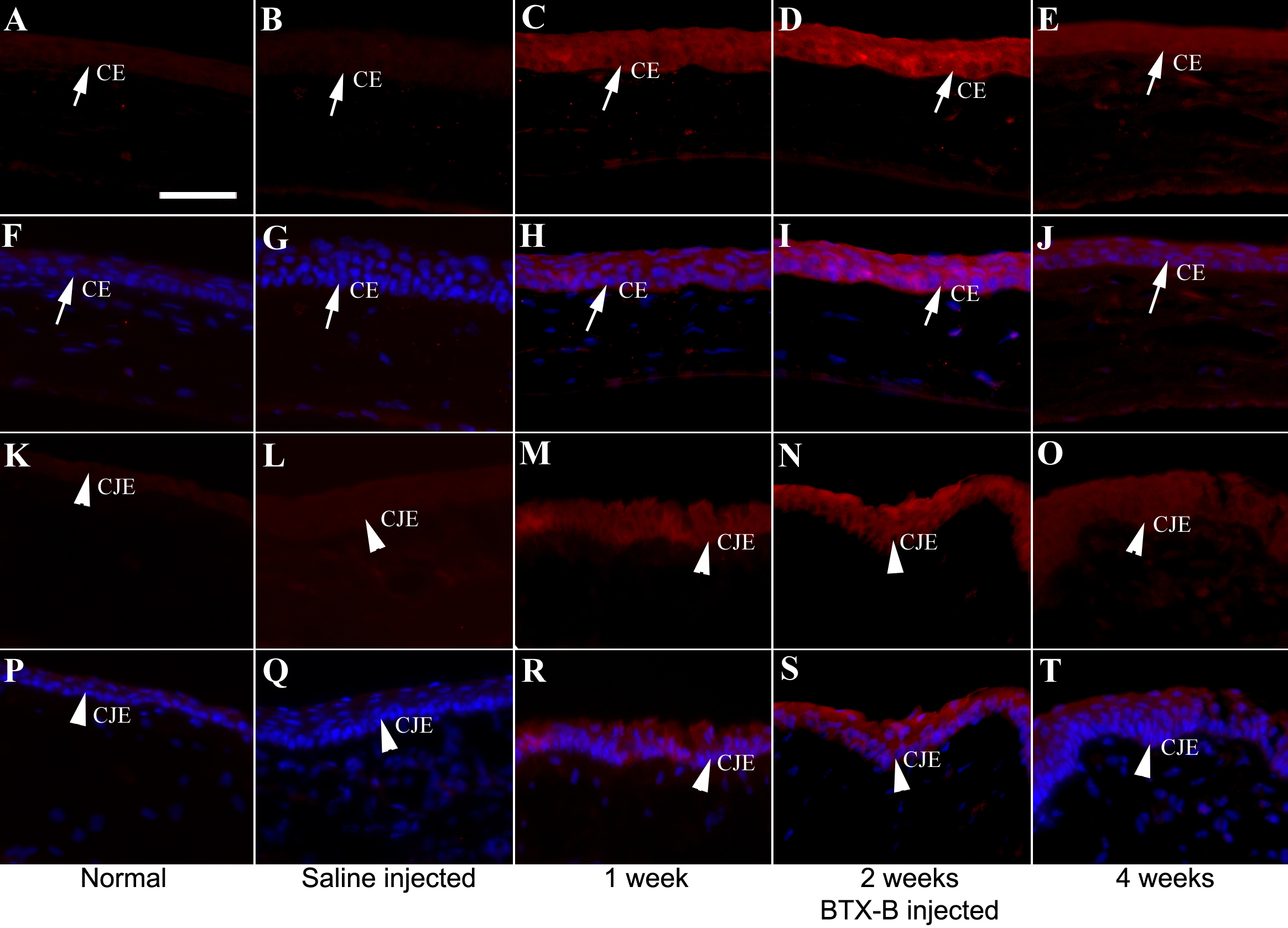
 Figure 6 of Zhu, Mol Vis 2009; 15:250-258.