Figure 6. Molecular analysis of gene expression levels after chemical cauterization in the untreated and bevacizumab treated mice. VEGF expression up-regulated in all time points, with a slight decrease following bevacizumab treatment (A). PEDF showed an inverse pattern of expression to VEGF (B). IGF-1 expression increased between days 8 and 10 following injury, with a higher level in the bevacizumab treated group (C). MIP-2 expression significantly increased in both the untreated and treated mice on day 2 and then dropped to near-normal levels
by day 14, with higher level in the bevacizumab treated eyes (D).
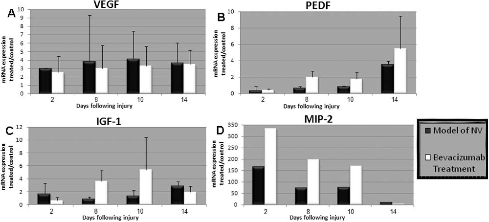
 Figure 6 of
Dratviman-Storobinsky, Mol Vis 2009; 15:2326-2338.
Figure 6 of
Dratviman-Storobinsky, Mol Vis 2009; 15:2326-2338.