Figure 3. Histology analysis using
hematoxylin-eosin staining. No blood vessels detected in the normal
cornea (A). Following cauterization, development of the scar
(arrow) was observed in the epithelial and anterior stroma of the
center cornea (B). New pathological blood vessels (C)
were located in the superficial stroma, filled with erythrocytes (*).
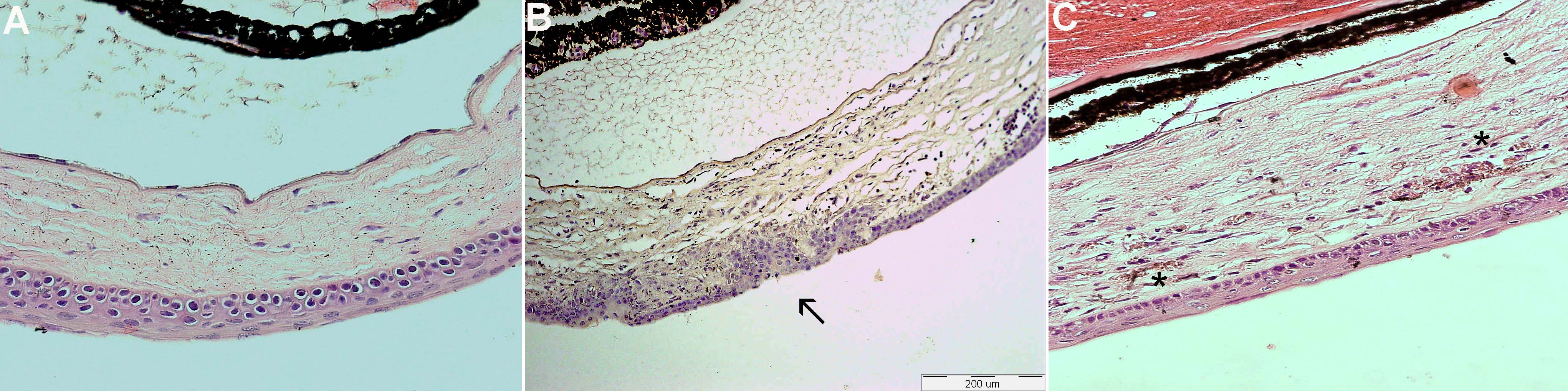
 Figure 3 of Dratviman-Storobinsky, Mol Vis 2009; 15:2326-2338.