Figure 2. Clinical appearance of
neovascularization in a mouse cornea. Comparison between normal eye,
without blood vessels on the cornea (A) and 8 days following
cauterization without treatment, with high level of neovascularization (B).
Less blood vessels observed after bevacizumab treatment by (H)
intravitreal injection, (I) intracameral injection or (J)
subconjunctival injection at the same time point. Fluorescein
angiography 8 days after chemical burn induction showing (C)
early leakage and (D-E) increased late leakage. Flat mount
cornea of the control mouse showing transparent cornea without blood
vessels (F). In the neovascular corneal model, studies with
fluorescein dye (G) and India ink (K, black) reveal new
vessels 8 days after induction of chemical burn, compared to the
control avascular cornea (K), and a reduced level of NV detected
following bevacizumab treatment (M-O).
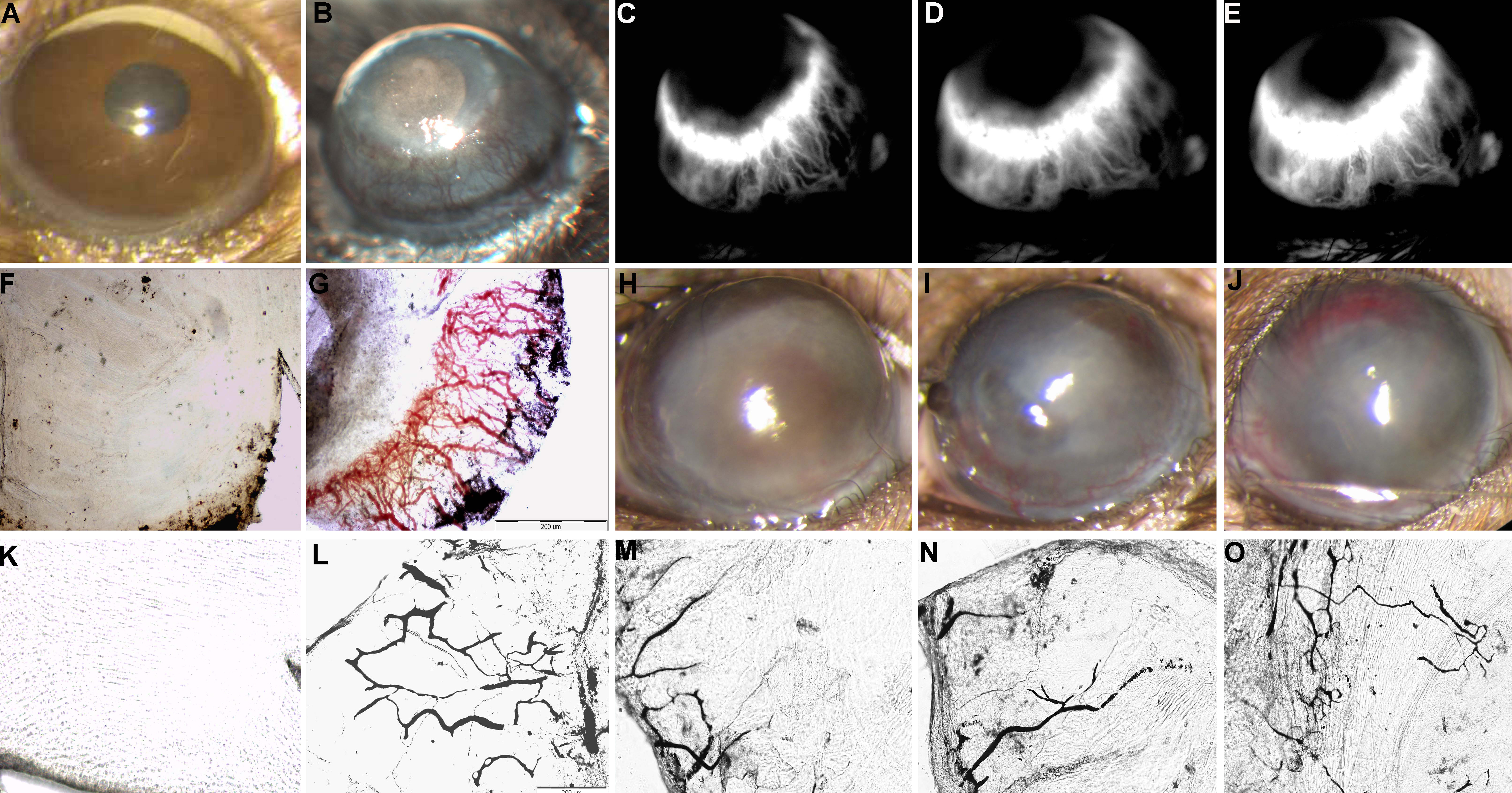
 Figure 2 of Dratviman-Storobinsky, Mol Vis 2009; 15:2326-2338.