Figure 1. Corneal neovascularization in
rabbits. A-B: Clinical appearance of neovascularization
in a rabbit cornea 10 days (peak) after chemical burn induction. Note
the central scar (yellow) covering approximately 10% of the corneal
area. C-D: Intracameral bevacizumab-treated cornea at 10
days. Note the reduced neovascularization.
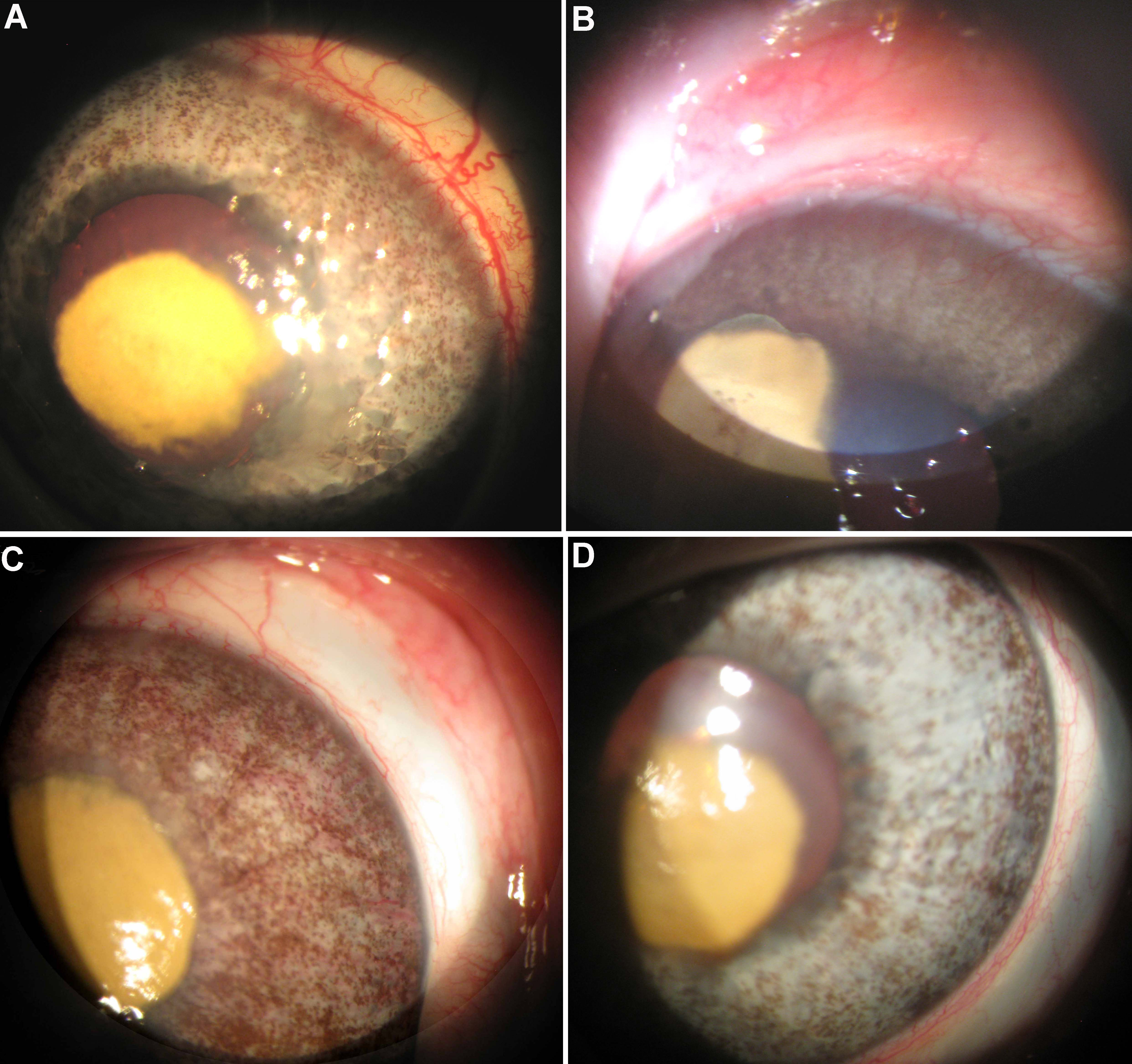
 Figure 1 of Dratviman-Storobinsky, Mol Vis 2009; 15:2326-2338.