Figure 7. ERG responses and fundus
photograph of Beagles with the RPGRIP1 insertion variant.
Bilateral scotopic (top) and photopic (middle) ERG recordings and
fundus photograph of the left eye (OS; bottom) of three Beagles: a 5y RPGRIP1+/−L
dog (A and D), a 2.8y RPGRIP−L/−L (B
and E), and another 2.8y RPGRIP1−L/−L dog (C).
Scotopic responses to a series of light stimuli are displayed with
increasing light intensity from top to bottom differing by 1 log cd/m2
up to 18,400 cd/m2. The photopic response was recorded with
31 Hz flicker stimuli of 35,900 cd/m2. Note the apparently
normal fundus appearance in the RPGRIP1−L/−L dog (E)
with undetectable cone response (B). OD indicates right eye.
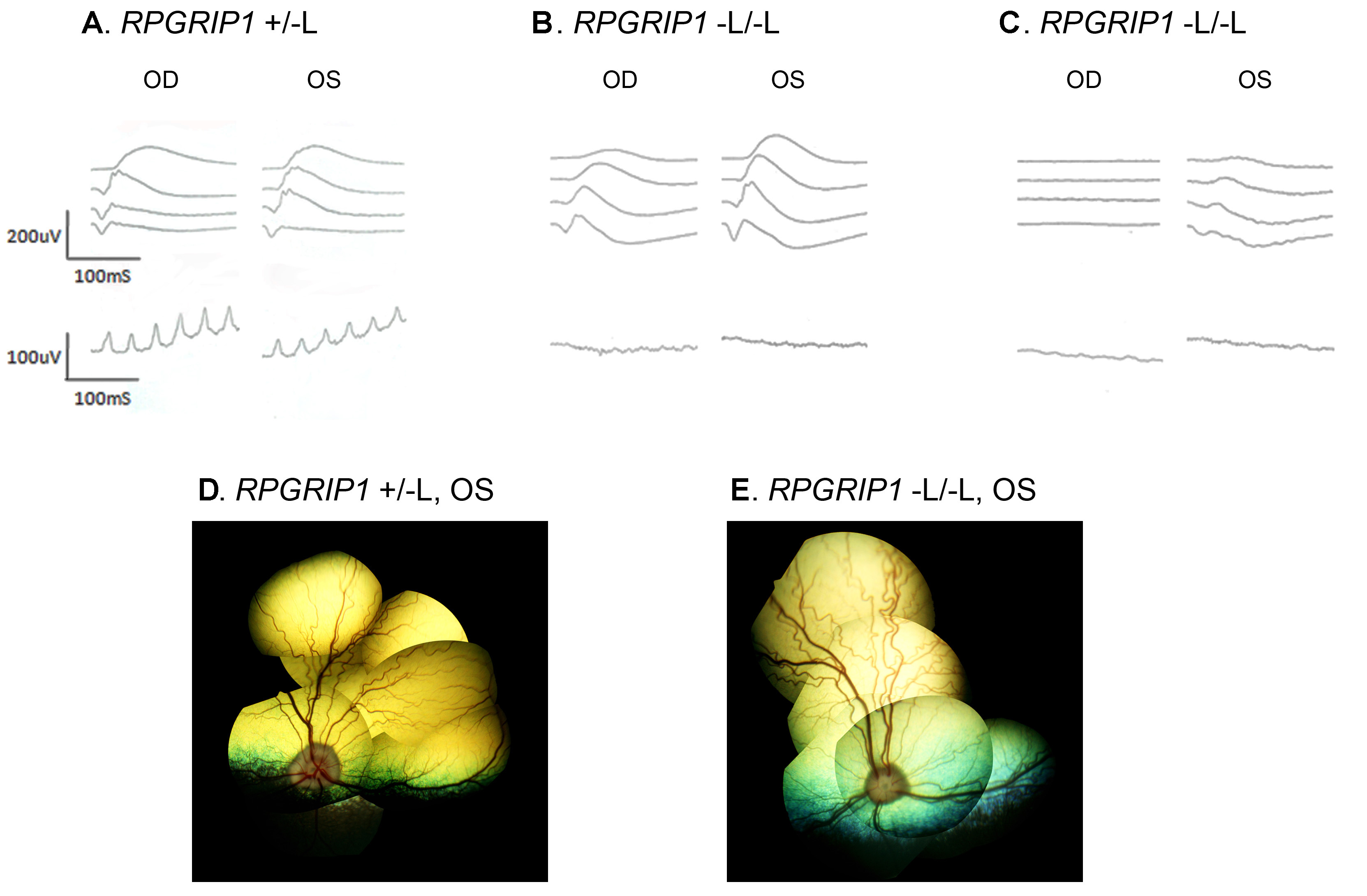
 Figure 7 of Miyadera, Mol Vis 2009; 15:2287-2305.