Figure 4. Immunoprecipitation of ERα followed by western blot analysis using anti-TERT antibody. Lane 1 is a non-immunoprecipitated
protein sample from normal LEC expressing minimal ERα. Lane 2 shows lack of interaction between ERα and TERT in normal canine
LEC. Lane 3 is a non-immunoprecipitated protein sample from cataractous LEC expressing more ERα than normal LEC in Lane 1.
Lane 4 demonstrates interaction between ERα and TERT in cataractous LEC. This experiment is representative of two repeated
experiments and was performed in reverse twice as well with similar results.
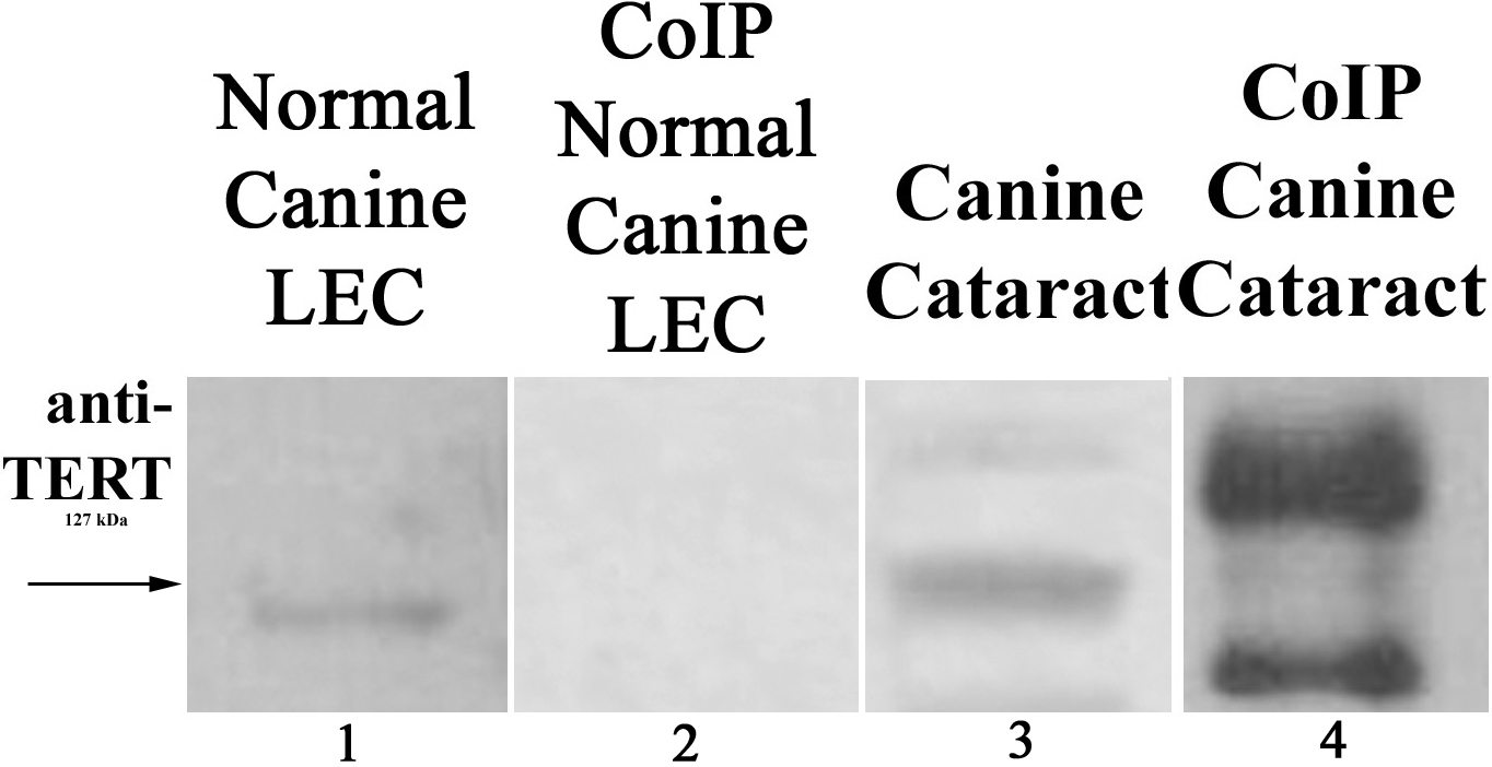
 Figure 4 of
Colitz, Mol Vis 2009; 15:2259-2267.
Figure 4 of
Colitz, Mol Vis 2009; 15:2259-2267.