Figure 2. Protein expression of ERα in normal and cataractous LEC. Representative results are shown of immunohistochemical staining
with anti-ERα antibody in normal canine LEC (A) and anterior capsulotomy samples from canine patients with naturally occurring cataract (B). B shows more extracellular matrix surrounding many of the cells, which is typical of LEC that have undergone EMT and make aberrant
collagens. Both the nuclei (large arrowheads) and the cytoplasm are immunopositive in the cataractous sample whereas the normal
sample has less intense immunostaining in both. Extracellular matrix production is seen in between and above the cataractous
LEC (red arrows). Immunostaining results are representative of repeated (three times) experiments. Chromogen is DAB with hematoxylin
counterstain. Magnification, 400×. C: Canine mammary adenocarcinoma shows nuclear and cytoplasmic immunopositivity in the acini and in some of the surrounding
stromal cells. D: Canine mammary adenocarcinoma served as a negative control and is representative of all negative control slides used. E: Western blot analysis compares the expression of ERα in normal LEC (three different age groups) and naturally occurring
breed-related/presumed-inherited cataractous LEC samples. Normal LEC express ERα less than the cataractous LEC. N=normal canine
lens. Controls include canine tumor samples overexpressing ERα (MCT=mast cell tumor, Mel=melanoma).
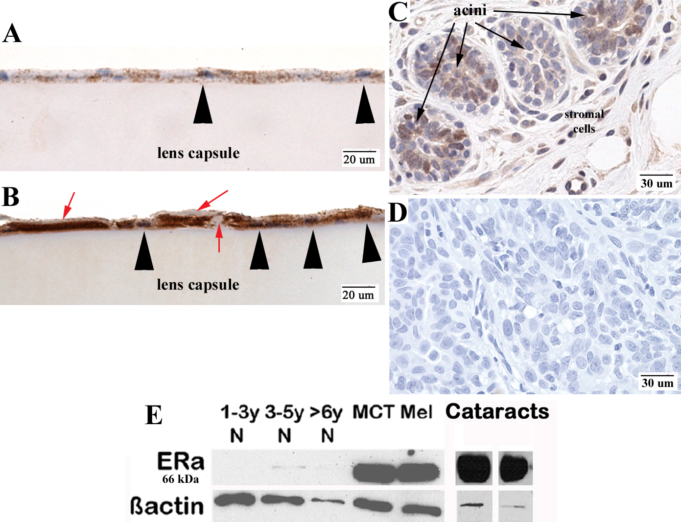
 Figure 2 of
Colitz, Mol Vis 2009; 15:2259-2267.
Figure 2 of
Colitz, Mol Vis 2009; 15:2259-2267.