Figure 3. Western blot analysis of
proliferating cell nuclear antigen, E-cadherin, Fibronectin-1, and MMP9
The results are representative of three independent experiments.
Western blot and densitometric analyses demonstrated that HCECs exposed
to AM (5% and 30%) evidenced increased levels of PCNA and MMP9 protein
(p<0.05 and p<0.001, respectively), whereas HCECs exposed to AM
(5% and 30%) exhibited reduced expression levels of E-cadherin and
Fibronectin-1 (p<0.05 and p<0.001, respectively). The values
displayed in these graphs are expressed as the means±SD of triplicate
determinations in a representative experiment. Values were normalized
to that of the actin expression.
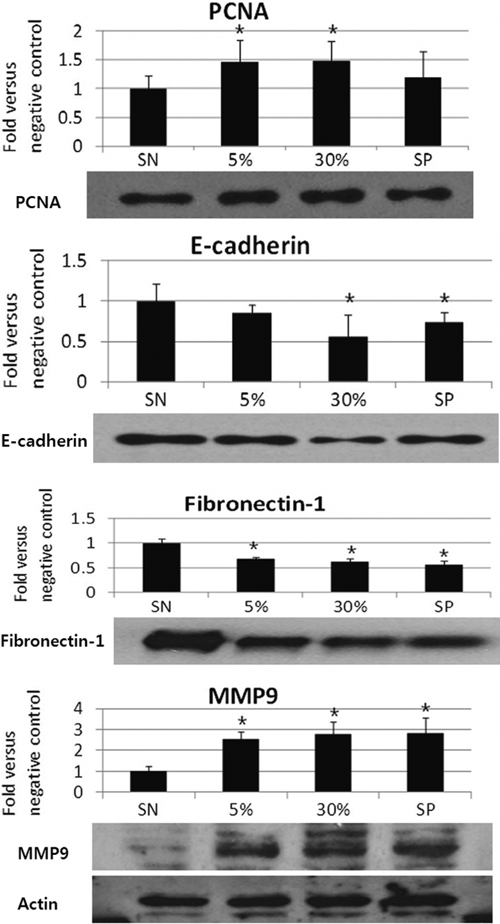
 Figure 3 of Choi, Mol Vis 2009; 15:2230-2238.