Figure 1. Migration assay by manual wounding of human corneal epithelial cells. HCECs were wounded by a yellow-tip in six-well plates.
A–D: Wounds in the negative control, amniotic membrane (AM) suspensions of 5% and 30%, and the positive control (SP) healed after
24 h. E: Statistically significantly increased healing rates, as compared to the negative controls were noted in the 30% AM, and
positive controls groups (asterisk).
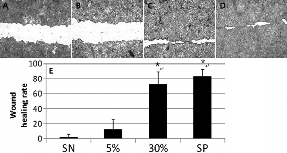
 Figure 1 of
Choi, Mol Vis 2009; 15:2230-2238.
Figure 1 of
Choi, Mol Vis 2009; 15:2230-2238.