Figure 1. Purified recombinant
crystallins. Recombinantly expressed proteins were purified to near
homogeneity as indicated by a single band on SDS-PAGE. Each protein (1
μg) was visualized with Coomassie blue stain on a 1.0 mm thick, 4-12 %
Bis/Tris gel. Proteins were βB1 (lane 1), βB2 (lane 2), βA3 WT (lane
3), βA3 DM (lane 4) and αA-crystallin (lane 5). Triangles indicate
positions of molecular weight markers.
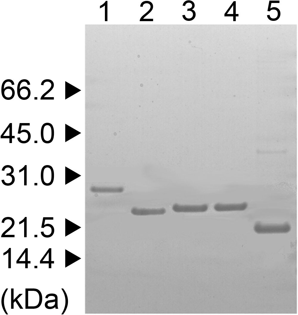
 Figure 1 of Takata, Mol Vis 2009; 15:241-249.