Figure 6. Cell lineage analysis by immunofluorescence. Double-labeling fluorescence with anti-CK7 (green) and anti-CD45 (blue) antibodies
was used to identify CK7+ secretory epithelial cells and CD45+ leukocytes, respectively. Moreover, propidium iodide (PI) dye
(red) was used to label nuclei. After 5 days of culture on denuded amniotic membrane (dAM) in 10% fetal bovine serum (FBS)
supplemented medium (FBSm10), all conjunctival cells showed green fluorescence (CK7+ cells) with red nuclei (PI). In contrast,
CD45+ cells were not observed in this culture (blue fluorescence). Scale bar 25 μm.
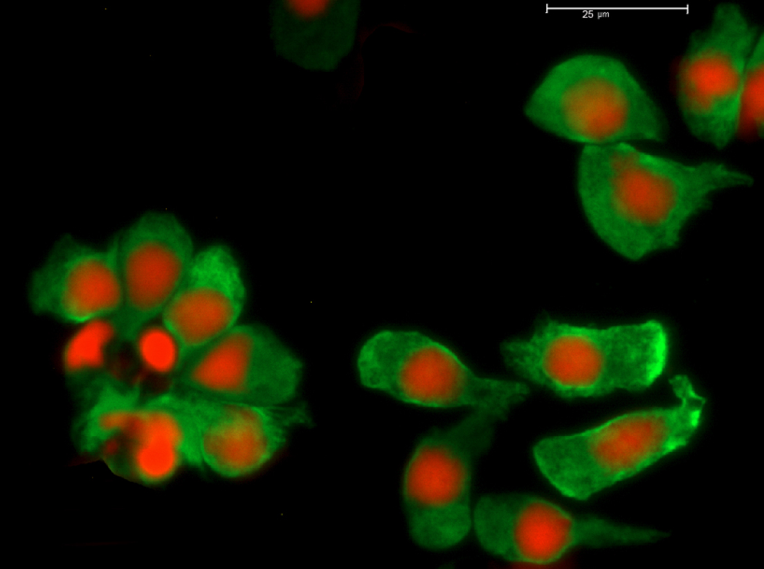
 Figure 6 of
Martínez-Osorio, Mol Vis 2009; 15:2185-2195.
Figure 6 of
Martínez-Osorio, Mol Vis 2009; 15:2185-2195.