Figure 4. Effect of media, substrata, and time. A: Multiple comparisons analysis showed statistically more living tarsal conjunctival cells on Day 5 with 10% fetal bovine
serum (FBS) supplemented medium (FBSm10) and denuded amniotic membrane (dAM) than the other media or substratum (p<0.011,
n=9). B: Temporal evolution of cultures showed that the highest number of living tarsal conjunctival cells was on Day 1 and decreased
(p<0.0001) on Days 2 and 5 (n=18), regardless of the substrate used. Values for A and B are means±95% confidence intervals.
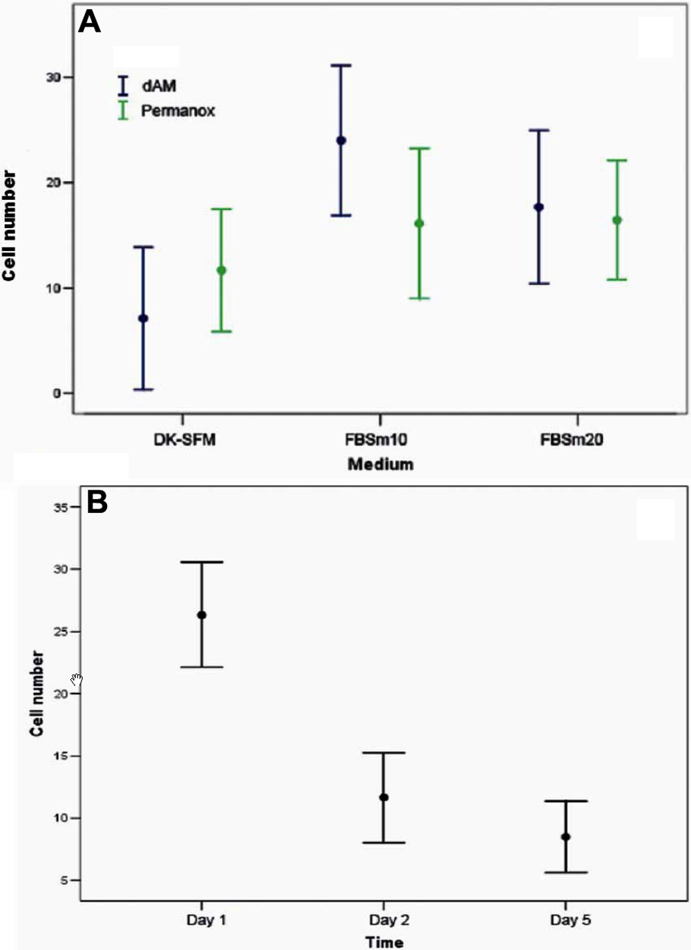
 Figure 4 of
Martínez-Osorio, Mol Vis 2009; 15:2185-2195.
Figure 4 of
Martínez-Osorio, Mol Vis 2009; 15:2185-2195.