Figure 2. Tarsal conjunctival cell
viability, apoptosis, and cell death determined by flow cytometry. A:
The morphological characteristics of recovered tarsal cells defined two
populations: larger, more complex cells (red) and smaller, less complex
cells (blue). Side scatter/forward scatter was used to define the
size/complexity of the cell pool. B: In this representative
experiment, early apoptotic cells (J4) and late apoptotic cells (J2)
composed 70.9% of the population of larger, more complex cells stained
with propidium iodide (PI) and annexin V, consistent with high level of
apoptosis. C: In a population of smaller, less complex cells,
76.5% (D3) had negative staining with PI and annexin V, indicating high
viability.
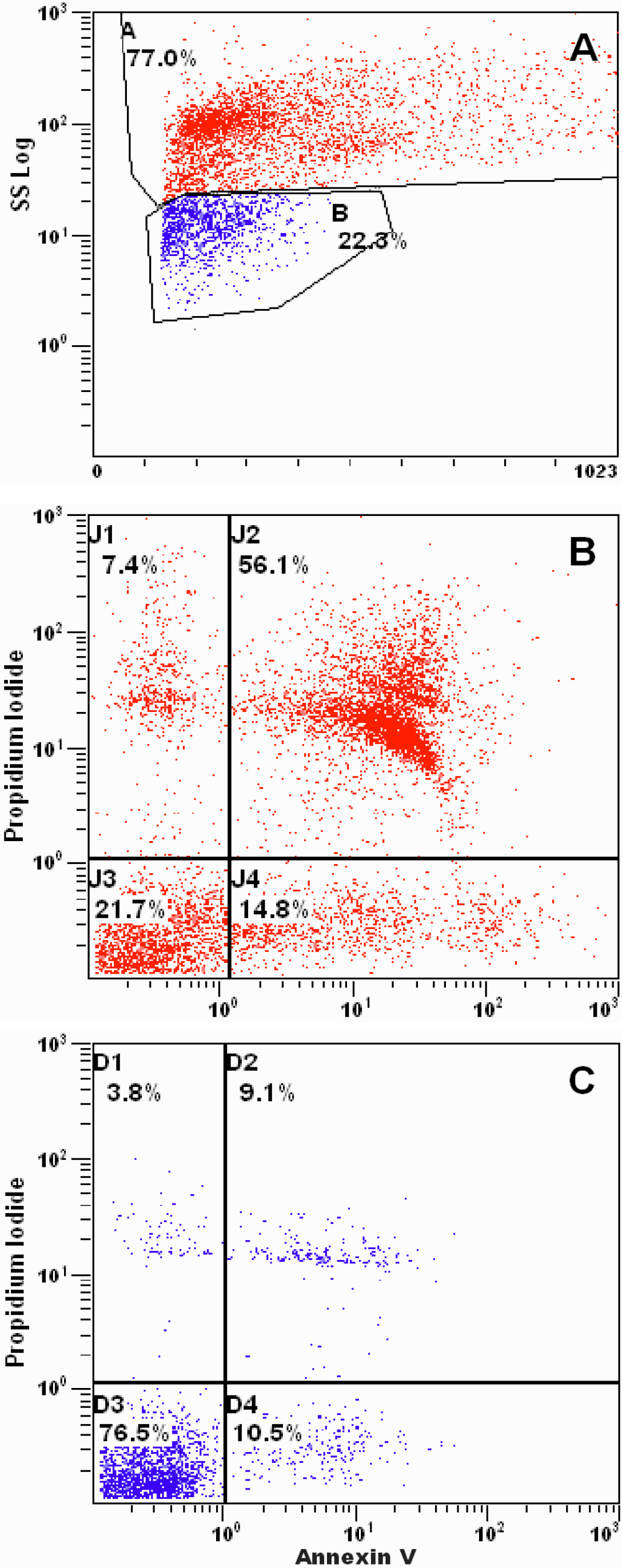
 Figure 2 of Martínez-Osorio, Mol Vis 2009; 15:2185-2195.