Figure 2. Subcellular localization of WT
and mutant NR2E3 proteins in COS-1 cells. COS-1 cells expressing WT or
mutant NR2E3 proteins were incubated with anti-Xpress antibody
(anti-Xp) and visualized using anti-mouse IgG-Alexa 488 antibody
(green). Central panels show nuclei labeled with bisbenzimide (blue).
The bottom panels display merged images. Scale bar represents 50 μm.
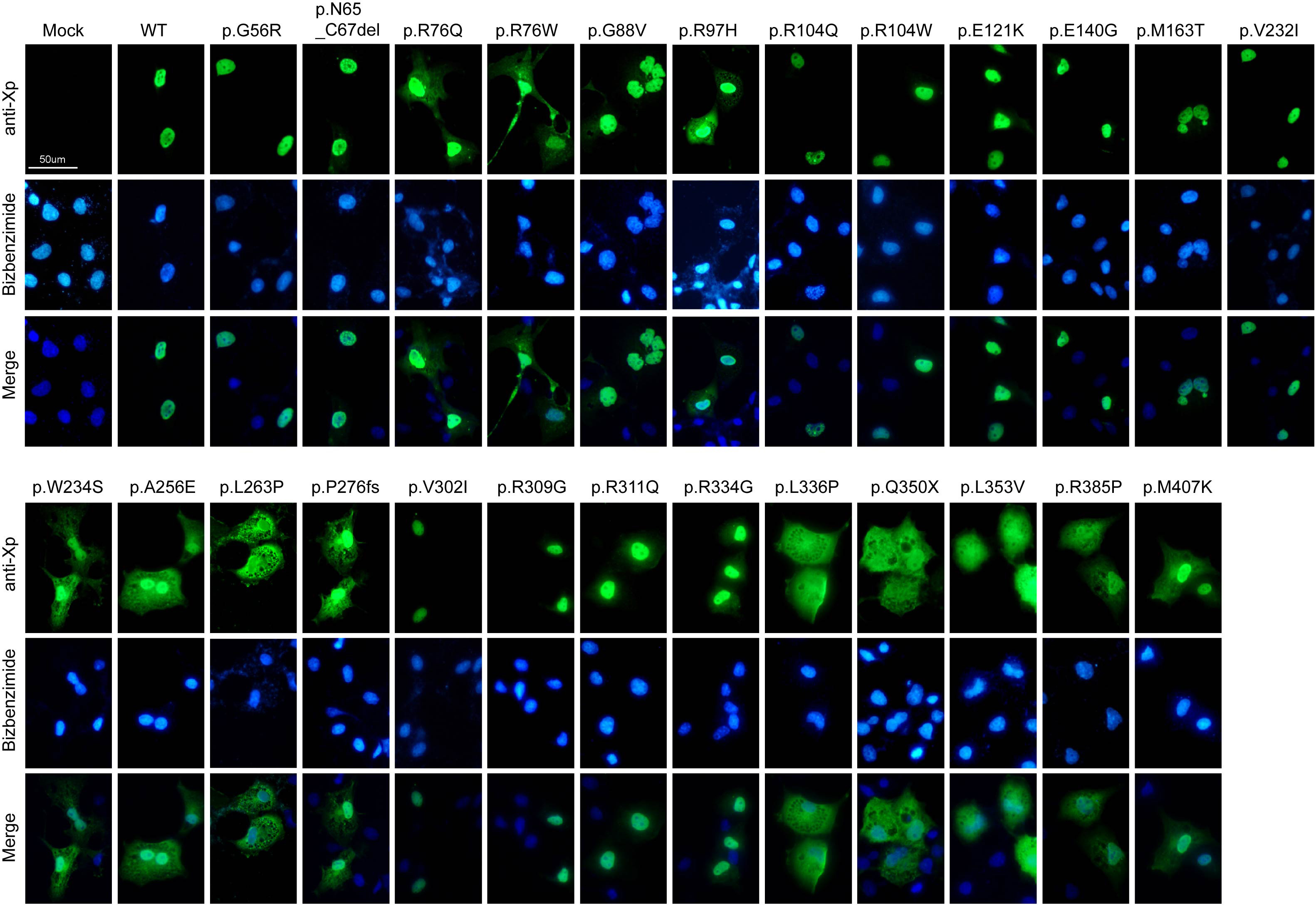
 Figure 2 of Kanda, Mol Vis 2009; 15:2174-2184.