Figure 1. Effects of galectin-1 on the morphology of cultured human retinal pigment epithelial cells and cell surface binding of galectin-1.
A-D: Human retinal pigment epithelial (RPE) cell suspensions were preincubated for 35 min without galectin-1 (A), or with 125 μg/ml galectin-1 (B), or 100 mM β-lactose before addition of 125 µg/ml galectin-1 (C), or 100 mM β-lactose (D) in the medium. RPE cells were then plated at a density of 0.5×10⁴ cells per well in 96-well plates and allowed to adhere for 90 min (A-D). The cells were observed by light microscopy (magnification 100×). E-H: Galectin-1 is detected on the surface of human RPE cells by immunofluorescence. Cells were cultured on glass coverslips
for 16 h before being treated with biotinylated galectin-1 (E). They were fixed, then stained with a fluorescent streptavidin conjugate. For controls (G), untreated cells were exposed to streptavidin conjugate alone. Nuclei were counterstained with 1 µg/ml Hoechst 33342 (F, H). Localization of bound galectin-1 was visualized by fluorescence microscopy at a 400x magnification. The bar represents
100 μm.
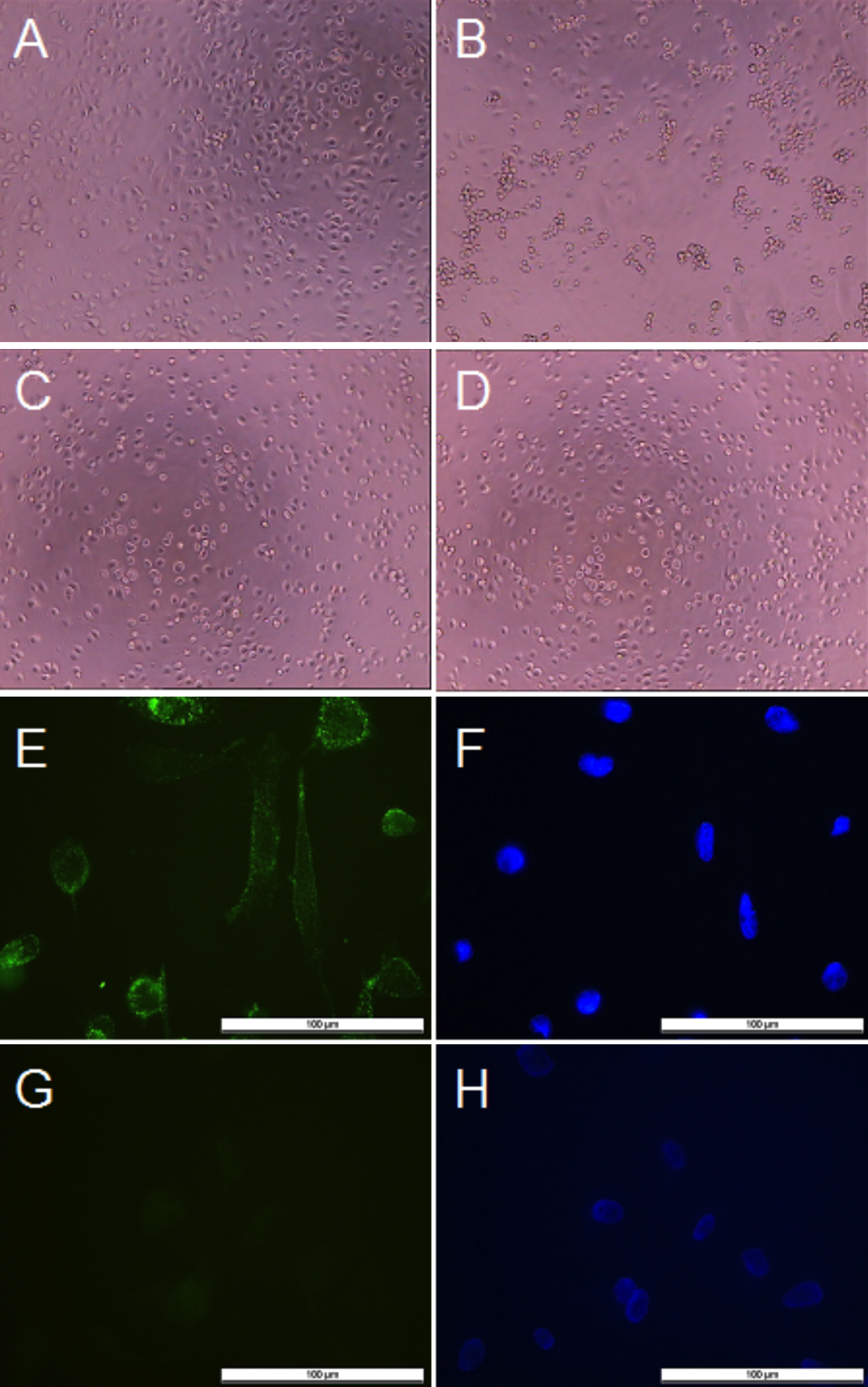
 Figure 1 of
Alge-Priglinger, Mol Vis 2009; 15:2162-2173.
Figure 1 of
Alge-Priglinger, Mol Vis 2009; 15:2162-2173.